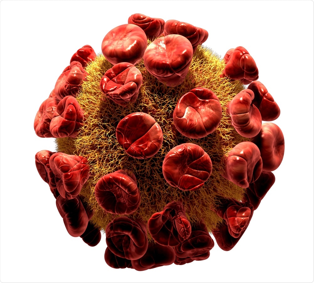
Due to their simultaneous expression, enveloped virus-like particles (VLPs) and extracellular vesicles (EVs) are difficult to distinguish and separate for analytical purposes. In addition to their co-expression, EVs and VLPs exhibit morphological, biophysical and molecular similarities.
To address this separation challenge, researchers recently devised a two-step chromatography method that can isolate VLPs and EVs for precise analysis.
 Ralwel | Shutterstock
One of the primary motives for developing a method to separate VLPs and EVs is to further our understanding of retroviral infections such as HIV. Although viral purification methods do exist, such as density gradient centrifugation, ultrafiltration or size-exclusion chromatography, these methods prove inefficient in their ability to separate EVs and VLPs.
The new technique is based on previous work utilizing heparin affinity chromatography as an alternative method to separate VLPs and EVs. Normally, high levels of heparin binding proteins within mammalian cell culture supernatants compete for binding sites in this single-step process.
The new two-step process was able to segregate the two particles without any competition. This research may pave the way for large-scale virology studies into the biological role of EVs and VLPs.
Methodology
Led by Dr. Alois Jungbauer, the researchers of this study developed a two-step chromatographic purification technique that begins with collecting both HIV-1 gag VLPs and EVs from HEK 293 cells. The cells were then treated and the supernatant collected and loaded into a column packed with core-shell beads.
The fluid fractions collected from the column were then further purified in a second step by heparin affinity chromatography. A second set of fluid fractions were then collected following the heparin affinity process and analyzed for total protein and double stranded DNA (dsDNA) content.
The analytical techniques used to separate, quantify and analyze protein content included Bradford assay, sodium dodecyl sulfate polyacrylamide gel electrophoresis (SDS-PAGE), Western blot analysis and enzyme-linked immunosorbent assay (ELISA).
Further particle characterization and size distribution analysis was performed through the use of nanoparticle tracking analysis (NTA) and transmission electron microscopy (TEM) imaging.
Results
EVs were found in significantly higher concentrations in the collected liquid fractions, whereas the VLPs were instead found in the elution peak. Not only was the new technique able to accurately separate the two molecules, it was also observed that VLPs and EVs maintained an intact spherical shape throughout the chromatography process.
Perspectives
Using this technique, the researchers were able to show that a two-step process can separate VLPs and EVs, allowing them to be studied in isolation. They are hopeful that this rapid and scalable technique can be applied to future large-scale production of VLPs and EVs to gain a deeper understanding into the biological function of these molecules.
Acknowledgements
The work performed in this study was supported by the Federal Ministry for Digital and Economic Affairs, the Federal Ministry for Transport, Innovation and Technology, the Styrian Business Promotion Agency SFG, the Standortagentur Tirol, Government of Lower Austria and ZIT - Technology Agency of the City of Vienna through the COMET-Funding Program, which is managed by the Austrian Research Promotion Agency FFG.
Source
Reiter, K., Aguilar, P. P., Wetter, V., Steppert, P., Tover, A., & Jungbauer, A. (2019). Separation of virus-like particles and extracellular vesicles by flow-through and heparin affinity chromatography. Journal of Chromatography A; 1588; 77-84.