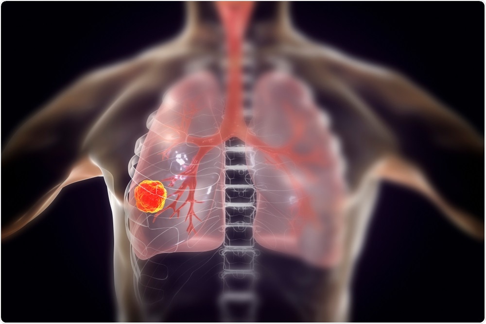
The researchers have been trying to teach the computer a deep learning algorithm for disease detection for some time. However, they have never been able to achieve such high rates of accuracy, until now.
 Kateryna Kon | Shutterstock
The team included 6,716 cases from the National Lung Cancer Screening Trial who assessed the efficacy of the algorithm. The success rates were high in accurate detection of the lung cancer cases from the screening results.
Another 1,139 independent clinical cases were also included in the experiment where the system was asked to analyse the results for detection of the cancers. Similar high accuracy was seen with both set ups when the AI deep algorithm was used.
The team conducted two studies. In one scenario they included cases where prior scans were available and in another these scans were unavailable. When the algorithm was presented with the lung CT scans it showed a greater accuracy compared to identification by six board-certified radiologists. When scans were not available the machine and the radiologists performed with similar accuracy. The machine scanned a total of 45,856 chest CT screens.
Pushing ‘the boundaries of basic science’
Dr. Daniel Tse, a project manager at Google in a statement said, “We have some of the biggest computers in the world. We started wanting to push the boundaries of basic science to find interesting and cool applications to work on.”
The whole experimentation process is like a student in school. We’re using a large data set for training, giving it lessons and pop quizzes so it can begin to learn for itself what is cancer, and what will or will not be cancer in the future. We gave it a final exam on data it’s never seen after we spent a lot of time training, and the result we saw on final exam — it got an A.”
Dr. Tse, Co-author
The team agrees that this is the first step and lot more needs to be done before the algorithm can be used on a routine bases for diagnosis. They explain that there are still problems of false positives – detecting cancer when there is none and false negatives or missing actual cases of cancer.
Dr. Eric Topol, director of the Scripps Research Translational Institute in California, in a statement said, “I’m pretty confident that what they’ve found is going to be useful, but it’s got to be proven.”
He was not involved in this study. Shravya Shetty, a software engineer at Google and a co-author of the study in a statement said, “How do you present the results in a way that builds trust with radiologists?” Showing them how the system works could help, she suggested.
The AI system uses 3D volumetric deep learning to analyze the full anatomy on chest CT scans, as well as patches based on object detection techniques that identify regions with malignant lesion.”
Dr. Shetty (Senior Author) and Dr. Tse (Co-author)
Dr. Mozziyar Etemadi, a research assistant professor of anesthesiology at Northwestern University Feinberg School of Medicine, one of the authors of the study, “It may start out as something we can’t see, but that may open up new lines of inquiry.”
Google researchers hope that this algorithm can be used in mass scale for screening and detection of lung cancer in large populations.
We are collaborating with institutions around the world to get a sense of how the technology can be implemented into clinical practice in a productive way. We don’t want to get ahead of ourselves.”
Dr. Tse, Co-author
Authors write that in 2018, lung cancers killed around 160,000 Americans. Screening for lung cancer can reduce the number of lung cancer deaths by 20 to 43 percent say the researchers and thus screening has been included in the US screening guidelines.
Dr. Topol said that AI would not replace radiologists but added, “It will make their lives easier. Across the board, there’s a 30 percent rate of false negatives, things missed. It shouldn’t be hard to bring that number down.”
If a radiologist makes a mistake, they can cause damage to one person while if the algorithm makes a mistake the damage may be to thousands of the population. Thus, the system needs to be rigorously tested before it is being used for mass screening.
This model will be available Google Cloud Healthcare API after the trials with the algorithm are tried with additional tests along with partner organizations.
Between hope, gloom, and perspectives
In a related research study, S. Heeke and colleagues from Nice University published a paper in the journal Annales de Pathologie last month, titled, “The age of artificial intelligence in lung cancer pathology: Between hope, gloom and perspectives.”
This team looked at the use of Artificial Intelligence in the histological analysis of the biopsy samples for the detection of lung cancers rather than radiological diagnosis using CT scans. Their review looked at the pros and cons of using AI in the pathological diagnosis of lung cancers using AI and its advantages and limitations as well.
Applying AI to medicine is a challenge
Authors WL Bi, Assistant Professor of Neurosurgery, Department of Neurosurgery, Brigham and Women's Hospital, Dana-Farber Cancer Institute, Harvard Medical School, Boston and colleagues published an article in CA: A Cancer Journal for Clinicians titled, “Artificial intelligence in cancer imaging: Clinical challenges and applications.”
The authors write, “Challenges remain in the accurate detection, characterization, and monitoring of cancers despite improved technologies.” They add that visual evaluations and interpretations using radiological images could be enhanced using computational programs. AI could help detect cancers faster and more accurately, they concluded.
For this study, the authors looked at the use of AI in four types of cancers such as lung, brain, breast, and prostate. They concluded that AI technology may soon be put to clinical use to “impact future directions in cancer care.”
Sources:
Ardila, D., et al. (2019). End-to-end lung cancer screening with three-dimensional deep learning on low-dose chest computed tomography. Nature Medicine. doi.org/10.1038/s41591-019-0447-x.
Heeke, S., et al. (2019). The age of artificial intelligence in lung cancer pathology: Between hope, gloom and perspectives. Annales de Pathologie. doi.org/10.1016/j.annpat.2019.01.003.
Linda, W., et al. (2019). Artificial intelligence in cancer imaging: Clinical challenges and applications. CA: A Cancer Journal for Clinicians. doi.org/10.3322/caac.21552.