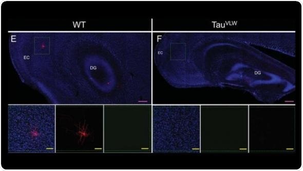
Dysfunctional neurons in the hippocampus of adult female mice modeling dementia can be repaired and reconnected to distant parts of the brain, reports a new study published in JNeurosci. The similarity between the mouse model and the human condition underscores the therapeutic potential of targeting these cells in dementia patients.

Representative images of the hippocampus and Entorhinal cortex. Credit:Terreros-Roncal et al. JNeurosci (2019).
The hippocampus generates new brain cells throughout life and is implicated in neurodegenerative diseases. María Llorens-Martín and colleagues at the Centro de Biología Molecular "Severo Ochoa" (CBMSO, CSIC-UAM) used a mouse model of frontotemporal dementia to investigate the effects of the disease on dentate granule cells.
Compared to control subjects, the researchers observed strikingly similar alterations in newborn neurons from their mouse model and from human brain tissue of patients with frontotemporal dementia. In mice, chemically activating the cells and placing animals in a stimulating environment with running wheels and toys reversed the alterations and restore some of the connectivity disrupted by dementia. If translated to humans, these results suggest potential new directions for combating cognitive decline in the elderly.
Source:
Society for Neuroscience
Journal reference:
Terreros-Roncal, J. et al. (2019) Activity-dependent reconnection of adult-born dentate granule cells in a mouse model of frontotemporal dementia. JNeurosci. doi.org/10.1523/JNEUROSCI.2724-18.2019.