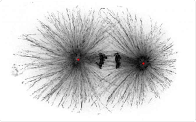
All life on earth depends upon the process of mitosis or cell division in which the chromosomal content of a cell is equally divided between two new cells. For mitosis to occur, the chromosomes in each pair must be pulled apart to opposite poles of the dividing cell. This action centers around the small cell organelles called centrioles which are prominent in all dividing cells.
Scientists at the Max Perutz Labs, a joint venture of the University of Vienna and the Medical University of Vienna, have examined how centrioles contribute to this process. The findings, published in "Developmental Cell", help to explain the function of centrioles in mitosis.
The centrioles form a dense network of radial proteins around them, called the pericentriolar material (PCM). This assembly forms a structure called the centrosome in a process called centrosome maturation. The centrosome comprises a filamentous spindle which must have an anchoring point to operate on the chromosomes. This anchoring point appears to be the centrioles themselves.
The PCM increases rapidly at the beginning of mitosis, and plays a fundamental role in organizing microtubules into the mitotic spindle filaments. Various proteins called PLK-1 and Aurora A/AIR-1 participate in this process.

Using laser microsurgery, they were able to remove the centrioles from within the centrosome at different stages of mitosis without destroying the entire structure. Image Credit: Alexander Dammermann
The fibers on either side of the mitotic spindle must be equal in size. Scientists suggest that this cannot be achieved by autocatalytic growth of the PCM fibers alone, but centriolar catalytic activity is also needed to keep growth on both sides equal. The centrosome is thus both the origin and the site of attachment for these fibers to operate in chromosomal segregation.
Though centrioles are vital to initiating centrosome formation, scientists don’t know how they contribute to further functioning of the PCM throughout the process of mitosis. This question was partly answered in the current research which used a model organism called C. elegans. The advantage of this organism is its very large centrosomes.
The use of laser microsurgery allowed for exceptionally precise cuts to be made in the dividing cells at various stages to remove the centrioles without disrupting the PCM or centrosome as a whole, so that the effects on the PCM could be directly observed.
The results of early centriole removal were that the PCM stopped growing, and indeed disintegrated as normally occurs at the completion of mitosis. Centrioles are thus essential for initiating PCM assembly.
During the phase of PCM expansion, centriole removal led to a halting of centrosome growth. The PCM already formed remained intact. However, there was marked loss of strength in the mitotic spindle as shown by the disintegrative effect of microtubule-mediated pulling forces. This was found to be because of the forces exerted by the migrating chromosomes via the microtubules on a weakened PCM. When the microtubules were broken down, the PCM remained intact. However, the loss of PLK-1 activity led to the disappearance of PCM. Thus PLK-1 alone is needed for PCM maintenance in mitosis, but when pulling forces are applied, centrioles are essential to maintaining the strength of the PCM.
However, when the centrioles were removed late in the cycle, in prometaphase, the PCM continued to stay in place and the current cycle of cell division and chromosome separation was completed. However, without centrioles the next cycle failed to show centrosome formation.
Centrosomes that had the centrioles removed showed signs of being on the verge of splitting apart under the stress of the opposing movement of chromosomes in each pair to the poles of the cell. Thus centrioles were found to confer structural integrity on the centrosome apparatus, despite the fact that the PCM is about 30 times larger than the centrioles themselves.
The researchers think that this function might be made possible by the centrioles acting as attachment sites for proteins which increase the tensile strength of the PCM, enabling it to withstand the pulling of the chromosomes. This would mean these tensile proteins acted somewhat like the steel rebars in reinforced concrete.
Study authors Triin Laos and Gabriela Cabral said, “What we found was that centriole ablation did not lead to an immediate collapse of the PCM as we had expected. However, further growth was strongly impaired, revealing a critical role for centrioles in PCM accumulation, and therefore mitotic spindle assembly.”
The study shows the vital role of centrioles in regulating the formation of PCM and its growth throughout mitosis, as well as ensuring its structural unity. However, mystery surrounds the actual mechanisms by which these minute organelles achieve such different functions.
Journal reference:
Gabriela Cabral, Triin Laos, Julien Dumont, Alexander Dammermann, 'Differential Requirements for Centrioles in Mitotic Centrosome Growth and Maintenance', Developmental Cell, 2019, https://doi.org/10.1016/j.devcel.2019.06.004, https://www.sciencedirect.com/science/article/pii/S1534580719305222