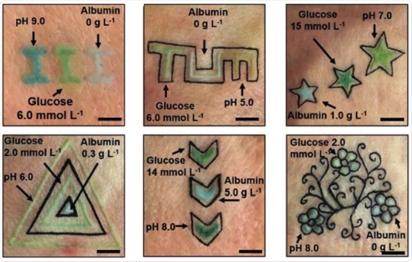
Scientists in Germany have developed an intradermal tattoo that changes color in response to fluctuations in glucose, albumin, and pH levels. Tests on animal skin have shown that the tattoos successfully change color when concentrations of key biomarkers were changed, marking an exciting step in real-time monitoring of chronic diseases like diabetes.
These sensors included a pH tracker consisting of methyl red dye, bromothymol blue dye, and phenolphthalein dye, which could alert doctors to cases of acidosis (low blood pH) or alkalosis (high blood pH), two conditions that are caused by a range of health problems.
The second and third sensors charted the levels of glucose and albumin (a carrier that transports protein in the blood). As is widely known, high levels of glucose can indicate cases of diabetes, while high levels of albumin can indicate heart problems, and low levels indicating liver or kidney failure.
The glucose sensor was made from the enzymatic reactions of glucose oxidase and peroxidase, and, when glucose levels changed, the color of the sensor changed to dark green or yellow. The albumin sensor, which turned green in response to changes in albumin levels, was made from a yellow dye.
The paper explained the function of these remarkable biosensor tattoos, stating they were “minimally invasive”, and that diagnostic devices that can continuously monitor metabolites in humans will “transform personalized medicine”, with patients living with chronic diseases and elderly people benefiting from continuous monitoring devices in particular.
 Credit: Yetisen et al., Angewandte Chemie International Edition, 2019.
The tattoos have not yet been tested in humans, with the tests being carried out on samples of pig skin. The color changes were evaluated with a smartphone camera and an app, with an algorithm determining biomarker concentrations by comparing chromatic variations of the biosensors to calibration points.
Calibration charts could be used to evaluate the color changes in different light conditions, including brightness, saturation, and shades.
“Body modification by injecting pigments into the dermis layer is a custom more than 4000 years old,” the researchers stated, but they recognized that damaging the epidermis (as is necessary to injecting pigment into the skin) may not always be desirable.
They suggested that microjet injections can pigment the skin without “sacrificing” the epidermal layer by injecting the pigment through electrical- or laser-induced shockwaves.
The authors write:
Here, a functional cosmetic technology was developed by combining tattoo artistry and colorimetric biosensors. Dermal tattoo sensors functioned as diagnostic displays by exhibiting color changes within the visible spectrum in response to variations in pH, glucose, and albumin concentrations.”
“The applications of the sensors can be extended to the detection of electrolytes, proteins, pathogenic microorganisms, gases, and dehydration status,” they continued, also adding that the tattoos may also find use in medical diagnostics to keep track of a “broad range” of metabolites.
Journal reference:
Yetisen, A. K., et al. (2019). Dermal Tattoo Biosensors for Colorimetric Metabolite Detection. Angewandte Chemie (International Edition). https://doi.org/10.1002/anie.201904416.