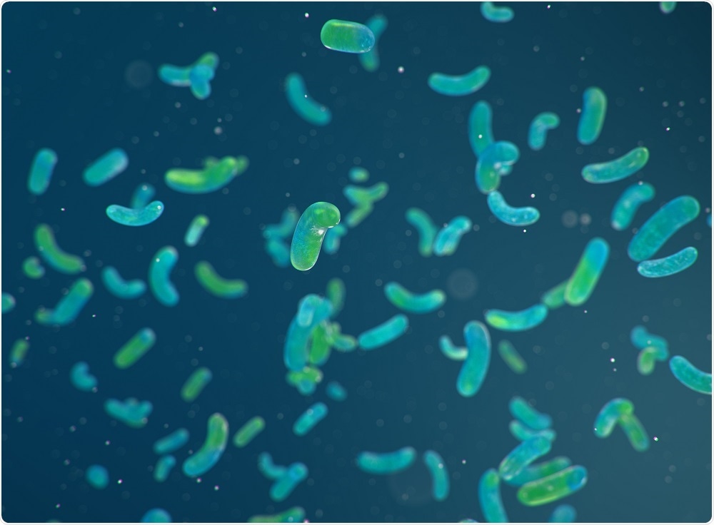
Some types of bacteria kill other cells by releasing proteins called “pore-forming toxins” (PFTs) that create holes in the cell membrane. The PFTs bind to the cell membrane and burrow into, creating a tube-like channel or pore. Once enough holes have been punched into the cell membrane by these toxins, the target cell destroys itself.
However, PFTs have captured the interest of scientists in many areas other than a bacterial infection. The nano-sized pores or nanopores that the toxins form can be used to “sense” biomolecules. A biological molecule such as DNA or RNA moves through the pore and its components (such as the nucleic acids in DNA) emit distinct electrical signals that researchers can detect.
 Ustyna Shevchuk / Shutterstock.com
Researchers study a major PFT called aerolysin
As reported in the journal Nature Communications, a team of researchers led by Matteo Dal Peraro at Ecole Polytechnique Fédérale de Lausanne has studied a key PFT called aerolysin that could enable more complex sequencing such as protein sequencing.
Produced by a bacterium called Aeromonas hydrophila, aerolysin is a key member of a major family of PFTs that is found across a wide range of organisms. A key advantage of the aerolysin toxin is that it creates very narrow pores that can differentiate between molecules at a much greater resolution, compared with other PFTs.
Researchers have previously shown aerolysin can be used to sense a number of biomolecules, yet few studies have yet explored the relationship between the toxin’s structure and its sensing capabilities.
Creating a computational model of the protein’s structure
To investigate, the team used computers to create a structural model of aerolysin that would help them understand how it's amino acids affect the overall function of the protein.
Once they had gained a basic understanding of this relationship, the researchers started to change various amino acids in the computational model. The model then went onto predict the potential impact these changes would have on the protein’s function.
Next, lead author of the study, Chan Cao, genetically engineered sixteen “mutant” aerolysin pores, which were embedded in lipid bilayers to mimic their position in the cell membrane.
Various measurements including molecular translocation experiments and single-channel recording were then carried out to investigate molecular regulation of the pore’s ionic conductance, ion selectivity, and translocation properties.
They found what drove the function between structure and function
This approach finally revealed what it is that drives the relationship between structure and function, which is the protein’s cap.
As well as the pore being a transmembrane tube-like channel, it also possesses a cap-like structure that binds a target molecule and pulls it through the channel. The researchers found that this process is dictated by electrostatics at this cap region.
The team will now be able to engineer customized pores for sensing applications
Peraro says that through understanding the details of how the structure of the aerolysin pore relates to its function, the team will now be able to engineer customized pores for various sensing applications:
These would open new, unexplored opportunities to sequence biomolecules such as DNA, proteins and their post-translational modifications with promising applications in gene sequencing and biomarkers detection for diagnostics."
Peraro and team have already filed a patent for their work sequencing and characterizing the genetically engineered aerolysin pores.
Journal reference:
Cao, C., et al. (2019). Single-molecule sensing of peptides and nucleic acids by engineered aerolysin nanopores. Nature Communications. DOI: 10.1038/s41467-019-12690-9.