Image Credit: Photoprofi30 / Shutterstock
The brain is a hive of ceaseless and diverse functions blending to produce cognitive ability. This includes the ability to learn, to remember, to think and to make decisions based on assessments. However, even as humans gain experience and wisdom with age, cognitive decline hampers brain function in older people.
Aging is also accompanied by reduced immune function, causing older people to fall prey to infections more easily and to have higher levels of inflammation.
These two areas of reduced function may be linked. In the current study, scientists found that the decline of activity in a set of immune cells called group 2 innate lymphoid cells (ILC2s) seems to be related to aging, and this is also linked to cognitive decline.
ILC2s are localized to specific tissues and are involved in repair processes following injury. A recent study showed that spinal LC2 cells enhanced healing following spinal cord injury. Their functions and locations are not completely known, however, as well as how they change with age.
The study
In the current research, the investigators looked at the brains of young and old mice to find if there were any ILC2s.
Increased number
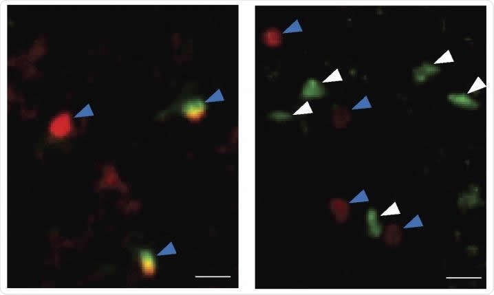
They found an age-related increase in the number of ILC2s in the choroid plexus, leading to up to 5 times as many cells in aged mouse brains. The choroid plexus is a highly vascular structure that secretes cerebrospinal fluid, the liquid that bathes the central nervous system. It is situated near the brain region called the hippocampus, which is known to be essential for memory and learning.
The researchers also looked at the brains of elderly humans and found that there were a lot of these cells accumulating with age. This is a sign of aging, as proved by the low number in younger mouse brains. They were mostly inactive.
Responsive to stimulation
However, they have a naturally long survival and a long period of activity, as seen in the next experiment. That is, when the researchers exposed them to a signaling chemical called IL-33, which is already known to stimulate ILC2s, this activated them to begin to proliferate vigorously and to synthesize the proteins that trigger the formation of new neurons as well as help them to survive.
Increased resistance
After the last dose of IL-33, they allowed a 4-week gap and then reassessed the number of ILC2s in both old and young mouse brains. They observed that over this period, the number of ILC2s In young mouse brains dropped to a fourth of the number present at the time of the last IL-33 administration. However, in the older mice, the activated ILC2s were still at about half the peak number. This shows that ILC2s in older mice are more tolerant of replication stress.

Staining for immune cells shows that the number of ILC2 cells (white arrows) are increased in the choroid plexus of old mice (right) compared with young mice (left). Other types of immune cells are indicated by blue arrows. Image Credit: Fung et al., 20200
More long-lived
The next experiment was aimed at evaluating the lifespan of the activated ILC2s in young and old mouse brains. They found that while both had the same proliferative capacity, the latter are more long-lived. They also seem to return to their quiescent state after the period of proliferation, when the stressor (IL-33) is withdrawn. This may account for their greater persistence following exposure to this cytokine, since quiescent cells suffer less DNA damage and other types of cell injury.
Responsive to secondary stimulation
However, the fact that their burst of proliferative activity died down after the stimulus was stopped did not affect their ability to be re-stimulated, as proved by a third experiment in which the aged mice were again treated with IL-33 for 2 days. This led to vigorous proliferation again. The scientists concluded that ILC2s in older mouse brains can switch between a state of dormancy and proliferation which allows them to live longer, which in turn is responsible for their gradual build-up with time.
Longer proliferation
Not only so, this self-renewing capability is greater in ILC2s from older mice, as they continued to multiply vigorously for 4 weeks without showing any features of exhaustion, in contrast to the two-week proliferation period of ILC2s from younger mice. They produce large amounts of functional proteins characteristic of these cells as well as of self-renewing stem cells.
Eventually, however, these cells begin to show signs of aging, but lymphocyte exhaustion marker proteins are reduced and remain low over time in these cells from aged mice.
Improved cognitive performance
Cognitive ability also improved in these treated mice, as well as in older mice treated with already activated ILC2s transferred into the cerebral ventricles, compared to ILC2s from younger mice. Researcher Kristen L. Zuloaga says, “This suggested that activated ILC2 can improve the cognitive function of aged mice.”
An interesting finding was that ILC2s from the choroid plexus in aged mice were more vigorous in their proliferative and functional renewal characteristics than those from the meninges, though both were at higher levels than those in younger mice.
Effects mediated by IL-5
When the researchers looked at the proteins secreted by activated ILC2s, they found that one of them was the cytokine IL-5. When old mice were treated with this chemical separately, their brains showed new nerve cells forming in the hippocampus, and a lower level of inflammation in the brain. This included a higher production of anti-inflammatory chemicals, a lower expression of pro-inflammatory cytokines, and a lower number of CD8 T cells that promote inflammation. The pro-inflammatory cytokine TNFα was also reduced, and its downstream signaling molecules. The number of and level of activation of the brain immune cells called microglia was also reduced. This probably leads a lower potential for damage. The scientists comment, “Together, IL-5 may directly act on aging-associated pro-inflammatory T cells to restrain neuroinflammation, which may lead to enhanced neurogenesis and improved cognitive function.”
Cognitive ability was found to be higher in this experimental group as well over a broad range of tests. “Together,” say the researchers, “our results highlight a novel role for IL-5 in alleviating aging-associated cognitive decline.”
Implications
In summary, the study concludes, “Our work has thus revealed the accumulation of tissue-resident ILC2 cells in the choroid plexus of aged brains and demonstrated that their activation may revitalize the aged brain and alleviate aging-associated cognitive decline.” The researchers think their work may lay the foundation for a new approach to preventing age-related damage to the brain that is responsible for several cognitive and degenerative disorders linked to neurologic injury.
Journal reference:
Fung et al. Innate lymphoid cells in the aged brain. Journal of Experimental Medicine. https://doi.org/10.1084/jem.20190915