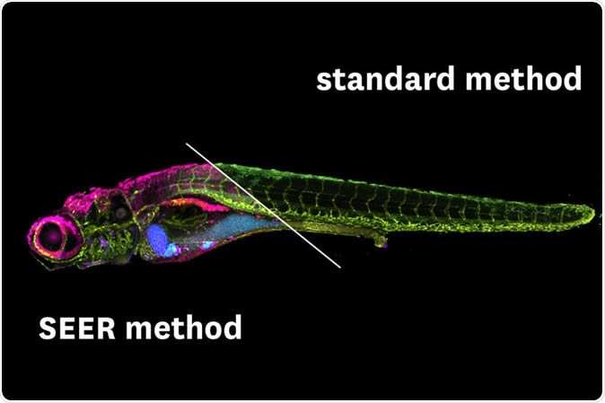
Scientists from the University of South California (USC) report the development of a novel technique to image living cells in greater detail and increased clarity. The study, published in the journal Nature Communications, describes a technology which can confer the ability to actually see things going wrong – a great advantage in the study of health and disease.

The new imaging technique is considerably faster and could even be used in a smartphone app for remote medicine or food safety. (Image/Wen Shi, Daniel E.S. Koo and Francesco Cutrale)
The basis
The foundation of the new technique is on the very basics of biological organisms. The problem with imaging living organisms at any level is the complexity of the processes that are taking place. Whether at whole-organism level or cellular level, proteins link different levels to each other, and it’s not clear to what level they belong, or what levels they actually connect.
The new imaging mode is based on fluorescence hyperspectral imaging (HSI). This method is advantageous for the following reasons:
- It distinguishes various colors
- It ‘sticks’ a tag onto molecules of interest to follow them through the imaging process
- It yields highly colored images of the internal workings of the organism
The issue with HSI
However, its use has been limited by the following shortcomings:
- It cannot pick up all the colors of the full spectrum
- It needs a tremendous amount of data because of the intricate nature of the system, which prolongs the time required for image acquisition and processing
- It produces the final digital image by complex and time-consuming calculations, which prevent real-time experimentation on the biological system.
This final limitation is very important in the medical and experimental fields. While a potential or present danger is caught by the image, the time taken for analysis means that it is too late to do anything about it by the time the information is presented to the scientist or doctor.
Researcher Francesco Cutrale sums up the issue with this method: “There is a number of scenarios where this after-the-fact analysis, while powerful, would be too late in experimental or medical decision-making. There is a gap between acquisition and analysis of the hyperspectral data, where scientists and doctors are unaware of the information contained in the experiment.”
The solution
To overcome these limitations, the researchers did what scientists do best: they came up with another method, called spectrally encoded enhanced representations (SEER). Cutrale says, “SEER is designed to fill this gap.”
The benefits of SEER are obvious:
- It produces much clearer images.
- It can recognize and file fluorescent tags of any color across the full spectrum to provide more detailed images.
- It is up to 67 times faster because it uses mathematical computations to sift through and analyze the images acquired. This is based on a very adaptable algorithm developed by Wen Shi and Daniel Koo at the Translational Imaging Center of USC.
- It produces images at 2.7 times higher definition.
- It uses less computer memory which is a crucial advantage when dealing with Big Data, a given in modern bioscience and computational biology.
The researchers describe it as a “fast, intuitive and mathematical way” of understanding what is being seen even as the images are coming in and undergoing processing.
Applications
The first application of SEER will, fittingly, be in the area of medical research. The algorithm behind SEER will be adapted for the detection of two currently vexing clinical questions: how to identify lung cancer in the early (and curable) stages, and how-to pick-up signs that the lungs might be suffering damage from pollutants. The study will be carried out in collaboration with the clinicians at Children’s Hospital in Los Angeles.
Meanwhile, other experimental scientists have taken to using SEER to speed up their workflow and collect useful data more rapidly.
Not only so, any imaging technology is likely to reach the customer-at-large. These powerful tools are capable of functioning well on smartphones, where they can be installed as apps, to improve the quality of image production in a variety of situations.
Implications
The researchers recognize the importance of their invention, which could be the key to many highly improved diagnostic and therapeutic modalities, such as identifying the presence of lung cancer or damage from pollutants. The technology is flexible enough to adapt it to fit into the confines of an app for smartphones, which could allow its use in telemedicine. In addition, this feature makes it ideal for much wider applications including testing foods for safety or detecting counterfeit notes.
Source:
From detecting lung cancer to spotting counterfeit money, this new imaging technology could have many uses - https://news.usc.edu/165448/new-imaging-technology-lung-cancer-counterfeit-currency/
Journal reference:
Shi, W., Koo, D.E.S., Kitano, M. et al. Pre-processing visualization of hyperspectral fluorescent data with Spectrally Encoded Enhanced Representations. Nat Commun 11, 726 (2020). https://doi.org/10.1038/s41467-020-14486-8