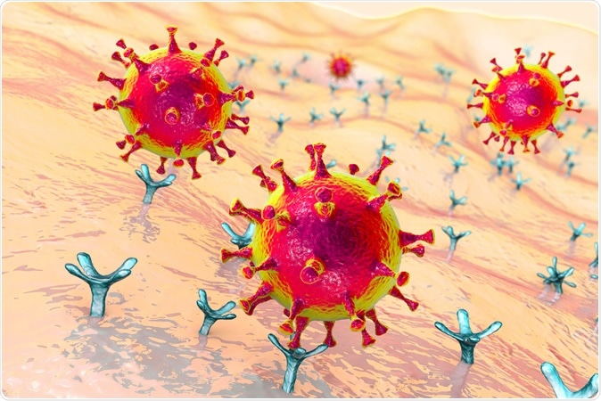
The world is still under the shadow of the COVID-19 pandemic, caused by SARS-CoV-2, a betacoronavirus which attaches to the angiotensin-converting enzyme 2 (ACE2) enzyme to enter and then infect the human host cell.

SARS-CoV-2 viruses binding to ACE-2 receptors on a human cell, the initial stage of COVID-19 infection, conceptual 3D illustration Credit: Kateryna Kon / Shutterstock

 This news article was a review of a preliminary scientific report that had not undergone peer-review at the time of publication. Since its initial publication, the scientific report has now been peer reviewed and accepted for publication in a Scientific Journal. Links to the preliminary and peer-reviewed reports are available in the Sources section at the bottom of this article. View Sources
The Importance of ACE2
The ACE2 is an enzyme that is generally found in the nasal epithelium, in the bronchial epithelium, and the type II pneumocytes. It is also found at other locations. ACE2 plays a part in other physiologic processes as well as in facilitating infection with SARS-CoV-2, to which it is considered by some to have more affinity compared to SARS-CoV.
ACE2 is a transmembrane protein with an extracellular domain containing 740 residues. A newer method of studying such proteins is surface plasmon resonance assay. When this was used to evaluate soluble ACE2 in bound form with the SARS-CoV-2 spike protein, it was found to have a high binding affinity falling in the range of nanomoles to picomoles. This property seems to confer potent antiviral neutralization activity.
Soluble ACE2
Soluble ACE2 has the ability to bind to the spike proteins of both SARS-CoV and SARS-CoV-2, preventing their entry into the cell. This protein has been used to treat patients with pulmonary hypertension and acute respiratory distress syndrome at dosages between 0.1 to 0.8 mg/kg, with a high tolerance limit.
The absorption, distribution, and taking up of soluble ACE2 is not likely to allow a prolonged period of viral neutralization in vivo. The molecule itself is not built for easy transport into the fluid over the epithelial lining of the lung from the bloodstream. Moreover, ACE2-IgG fusion proteins are known to bind the virus and neutralize the pseudoviruses carrying the SARS-CoV-2 antigens in vitro.
Some ACE2 variants that have mutations in the catalytic domain are also known to bind to and neutralize the virus. On the other hand, these fusion proteins can still retain FcRγ binding. This can destabilize the enzyme in serum or activate the FcRγ on the myeloid white blood cells, potentially causing a hyperimmune reaction in COVID-19, which could worsen the situation.
Mutant Fusion Proteins Neutralize Virus
The researchers made use of the higher affinity of the SARS-CoV-2 spike protein to the human ACE2 (hACE2) compared to the SARS-CoV to develop a therapeutic strategy that can block viral entry. This included introducing mutations in the catalytic domain of the ACE2 and in the IgG1 constant region, to prevent binding to the FcRγ receptor.
The researchers, therefore, generated four fusion proteins from ACE2 and IgG, all carrying the LALA mutation which prevents FcRγ binding and thus averts the potential danger of inappropriate immune reaction, while still allowing its binding to the neonatal Fc receptor. This allows it to be stable in serum while allowing transport into the lung tissue.
Apart from using the wildtype ACE2 extracellular domain, they also introduced several other mutations to disable the catalytic action of the protein on angiotensin, some of which are MDR503, MDR504, and a variant carrying two mutations, MDR505. The new fusion proteins were expressed as homodimers in transfected cells in culture, and bound to the monomeric RBD of the pseudotyped SARS-CoV-2 model, as well as the trimeric spike protein.
The hACE2-Fc constructs were examined for neutralization activity, and found that both MDR504 and MDR505 were more potent neutralizers than the wildtype hACE2-Fc IgG1, with a slightly less level of activity with the MDR503 form. At lower concentrations, both mutant forms were much more potent than the wildtype, and the MDR504 was found capable of inhibiting viral activity in 50% of the cultures.
The MDR504 Mutation Comes Out Ahead
The effect was most significant with the MDR504 mutation, which means it is an effective inhibitor of viral RBD or spike protein binding with the ACE2. How this happens is unknown, though cryo-electron microscopy shows that the RBD of the SARS-CoV-2 can bind the amine terminus of the hACE2 protein. Since the RBD also binds other residues, it is quite possible that this binding is the real target of the MDR504.
The highest binding was observed with the mutant MDR504 at room temperature and at 37 oC, which is in agreement with the increased virus neutralization seen in a cell plaque assay. Secondly, the MDR504 variant is as serum stable as the wildtype ACE2-Fc, yet appears at somewhat higher and therapeutically relevant concentrations in the lung lining fluid when injected into a mouse model.
The study concludes, “The MDR504 mutant appears to be an excellent candidate to move forward in terms of preventing or treating COVID-19.” Its utility as a preventive might be particularly high in people who have contraindications for vaccine use like blood cancer, or the use of immunosuppressive drugs.

 This news article was a review of a preliminary scientific report that had not undergone peer-review at the time of publication. Since its initial publication, the scientific report has now been peer reviewed and accepted for publication in a Scientific Journal. Links to the preliminary and peer-reviewed reports are available in the Sources section at the bottom of this article. View Sources