The results of a recent multinational research endeavor, currently available on the bioRxiv* preprint server, display the biological framework of SARS-CoV-2-related coronavirus in pangolins and reveal striking similarities to coronavirus disease (COVID-19) in humans, but also show that it can be transmitted vertically to the fetus.
The COVID-19 pandemic, caused by the severe acute respiratory syndrome coronavirus 2 (SARS-CoV-2), continues to reshape the entire globe. The number of cases worldwide is growing at an unprecedented pace, but so is the body of research in both fundamental and clinical aspects of the disease.
We are already well aware that the spike glycoprotein (S-protein) of SARS-CoV-2 binds to the angiotensin-converting enzyme 2 (ACE2) receptor responsible for cell entry. At the same time, the serine protease TMPRSS2 mediates S-protein activation and, in turn, assists viral invasion.
And due to a global spread of COVID-19, the potential risk of SARS-CoV-2 vertical transmission of SARS-CoV-2 has to be taken seriously. One of the reasons is that the ACE2 receptor is widespread in the placenta during pregnancy.
Moreover, SARS-CoV-2-related coronavirus (SARSr-CoV-2) that was previously isolated from Malayan pangolins shows close relatedness to SARS-CoV-2 – most notably in the receptor-binding domain (RBD) of the aforementioned S-protein.
"Understanding the biology of SARSr-CoV-2 in pangolins will be important for understanding key aspects of coronavirus disease", state researches from China, Australia, and Canada who decided to explore both viral and disease properties in a selected population of pangolins.
Do pangolins also get COVID-19?
In this study, the researchers used the viral gene that codes S-protein to recognize the infection with the pangolin-derived SARSr-CoV-2 and found that 15 animals in total – including six pregnant females – were naturally infected by the aforementioned virus.
In a nutshell, these Malayan pangolins that were positive for SARSr-CoV-2 exhibited respiratory symptoms such as cough, shortness of breath and dyspnea; the latter was indicated by the elevated levels of total carbon dioxide, partial pressure of carbon dioxide and serum bicarbonate.
Furthermore, computed tomography (CT) scan revealed diffused bilateral distribution of ground-glass opacities in the lungs, akin to the early phase COVID-19 pneumonia. Likewise, pronounced air bronchogram, transparent line under the pleura, and fibrotic streaks that were observed are similar to the advanced phase of COVID-19 disease.
In addition, the transcriptome analysis demonstrated an inadequate interferon response, with a plethora of dysregulated cytokine and chemokine responses in all animals (i.e., pregnant and non-pregnant adults and fetuses).
"Similar to human tissues infected with SARS-CoV-2 or SARS-CoV, a gene enrichment analyses illustrated that SARSr-CoV-2 triggered a dysregulated immune system in pangolins", study authors explain their findings in the bioRxiv paper. "Notably, non-pregnant adult, pregnant pangolins and fetuses had very different immune responses to the virus," they add.
Viral affinity for a variety of tissues
Generally, virus-positive pangolins exhibited increased expression levels of ACE2 and TMPRSS2 in all tissues in comparison to virus-negative ones. Clearly, the tissues with the highest levels of these cell-entry mediators also harbored SARSr-CoV-2 genetic material.
This supports the notion that cells expressing both ACE2 and TMPRSS2 are most amenable to coronavirus infection. Also, the study has shown that such cells are most pervasive in the liver, lungs and spleen when all of the tissues are mutually compared.
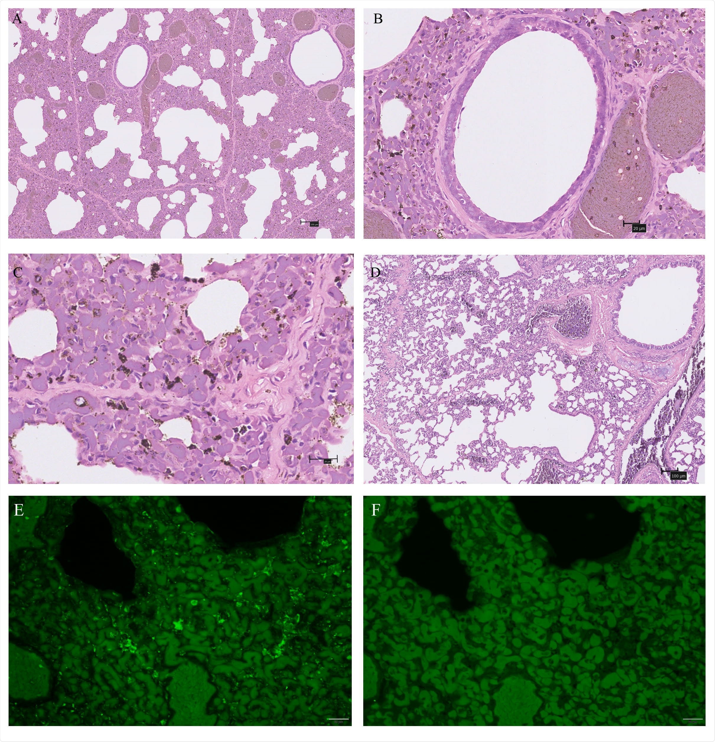
Nonetheless, SARSr-CoV-2 was also detected in the kidneys and intestines – tissues that do not have high expression of these receptor genes in the pangolins. Still, it must be emphasized that the lungs are the primary viral target.

Characteristic pathological changes and virus antigen expression in the lungs of one SARSr-CoV-2 positive pangolin . (A-C) An autopsy lung with SARSr-CoV-2 infection compared to a negative one (D) were stained by hematoxylin and eosin (H&E). (B) bronchiectasis; (C) the interstitium and the walls of the alveoli thicken. (E-F) Immunofluorescence for virus antigen expression in pneumocytes. (E) nucleocapsid of SARS-CoV-2 (Green); (F) negative control. Scale bars of (A) and (D) are 100um, while others are 20um.
The possibility of vertical SARS-CoV-2 transmission
Of the six pregnant pangolins positive to SARSr-CoV-2, in the three of their fetuses, the researchers detected RNA and nucleocapsid protein in the intestine, spleen, and muscles. This means that the vertical transmission of SARSr-CoV-2 occurring in utero is a realistic possibility.
But thus far, this finding cannot be translated to humans. Albeit neonatal cases of COVID-19 infection and antibodies in newborns have been found, testing was performed solely in newborns (rather than embryos); therefore, the horizontal transfer of infection after birth, or even the transfer of antibodies via placenta and breast milk, cannot be excluded.
In conclusion, this study shows comparable properties of SARSr-CoV-2 in pangolins and SARS-CoV-2 in humans – from disease presentation to tissue tropism and potential vertical transmission. Although further studies will be needed to corroborate these resemblances, pangolins could definitely serve as model animals in the interim.

 *Important notice: bioRxiv publishes preliminary scientific reports that are not peer-reviewed and, therefore, should not be regarded as conclusive, guide clinical practice/health-related behavior, or treated as established information.
Journal reference:
- Preliminary scientific report.
Li, X. et al. (2020). Pathogenicity, tissue tropism and potential vertical transmission of SARSr-CoV-2 in Malayan pangolins. bioRxiv. https://doi.org/10.1101/2020.06.22.164442.