A new study by researchers at Columbia University and the International Center for Infectiology Research, Inserm and published on the preprint medRxiv* in July 2020 reveals that children with the condition Multisystem Inflammatory Syndrome in Children (MIS-C) have a distinctly different pattern of antibody generation compared to adults with COVID-19 disease. This could provide a better understanding of both diseases and help the development of effective therapies based on the age group and symptoms.
With COVID-19 cases having increased immensely across the world, children were initially thought to be spared by the disease. Now, over six months since the emergence of severe acute respiratory syndrome coronavirus 2 (SARS-CoV-2) in China, pediatric COVID-19 is seen to be generally mild. However, a significant minority of children with COVID-19 present with some unusual symptoms.
One of these is MIS-C, a systemic inflammatory response seen in children infected with the SARS-CoV-2 virus, with apparent similarities to the vasculitic syndrome called Kawasaki disease or to the toxic shock syndrome. Affected children have symptoms relating to the inflammation of the gut, heart, lungs, kidney, brain, skin, or eyes. Though the syndrome is now well-recognized, the immune response is not so well characterized.
Anti-S Antibodies Important in COVID-19
The typical feature of antiviral immunity, induced by infections or vaccines, is the generation of specific antibodies. Similarly, people with active infection have been found to have specific anti-SARS-CoV-2 antibodies targeting the Spike or S protein. This is the primary protein antigen on the viral envelope that binds the host cell receptor, the angiotensin-converting enzyme 2 (ACE2). Anti-spike protein antibodies are therefore capable of neutralizing the virus, that is, of preventing viral entry into the host cell, and their use for therapy is being pursued for patients who are severely ill with COVID-19. Vaccine development is also following the ACE2 and S protein path.

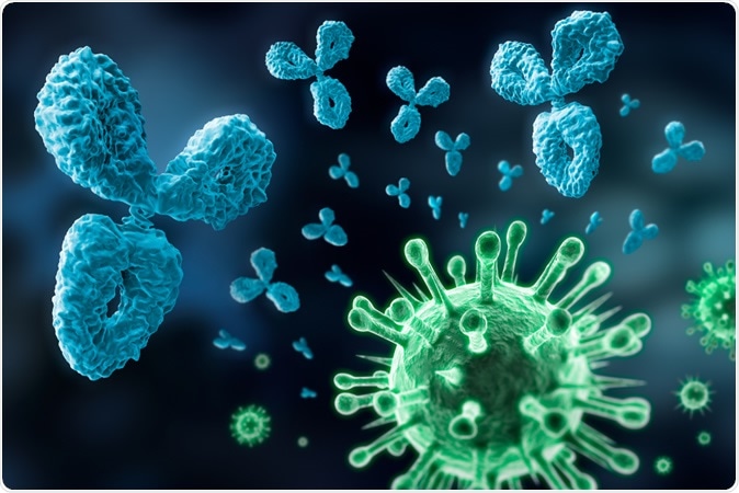
Antibody and virus - visual concept of the immune system. Illustration Credit: Peter Schreiber / Shutterstock

 This news article was a review of a preliminary scientific report that had not undergone peer-review at the time of publication. Since its initial publication, the scientific report has now been peer reviewed and accepted for publication in a Scientific Journal. Links to the preliminary and peer-reviewed reports are available in the Sources section at the bottom of this article. View Sources
The Study: What Antibodies, How Much, and What Do They Do?
The current study examines the antibody response concerning its specificity and functionality, as well as its ability to protect the individual against infection. The study was carried out in three groups of patients at the peak of the pandemic in New York, during the period March to June 2020. This includes patients who had a mild form of the infection and recovered, and who were asked to donate convalescent plasma. It also includes hospitalized patients with critical COVID-19, namely, acute respiratory distress syndrome (ARDS), and children with MIS-C.
The COVID-19 patients were of various ages from 14 to 84 years, while those with MIS-C were between 4 and 17 years. Adults with COVID-19 had other comorbidities but not the children with MIS-C.
Systemic Inflammation Common to MIS-C and COVID ARDS
All samples were collected from hospitalized patients within 36 hours of admission or intubation. The period from clinical symptoms to sample collection was shorter with MIS-C than with COVID-19 patients who developed ARDS. Blood levels of pro-inflammatory markers were much higher in both sets of patients, indicating systemic inflammation. These include IL-6 and C-reactive protein (CRP).
On the other hand, ferritin and lactate dehydrogenase (LDH) levels are much higher in COVID patients with ARDS than with MIS-C patients. Moreover, ARDS never occurred in the MIS-C cohort, who had less severe organ damage compared to the COVID-19 group. This suggests that the infection in the two groups followed different inflammatory and systemic pathways.
IgG in MIS-C, All Subtypes in COVID ARDS
The researchers first looked for binding of plasma IgG antibodies to recombinant spike protein on the cell surface, to confirm that the two disease manifestations are linked to the presence of specific anti-SARS-CoV-2 antibodies. They found that in all patients, but not in control samples collected before the pandemic began, the S protein and its common D614S variant was bound by plasma antibodies, which failed to bind SARS-CoV or MERS-CoV S protein.
They then analyzed the antibodies by the specificity of binding and antibody class. They found that IgM is first produced in the primary immune response, followed by IgG and IgA in plasma and in secretions, respectively. They found high levels of IgM, IgG, and IgA antibodies targeting the S protein in both COVID patients and convalescent plasma donors (CPD), compared to negative controls. The highest antibody level was found in severely ill COVID-19 patients with ARDS.
On the other hand, anti-S antibodies in children with MIS-C were mostly IgG, with some IgA, at levels relative to those in the CPD. On the other hand, IgM levels in these patients were low, comparable to those in control plasma. The IgG to IgM ratio was higher in the MIS-C cohort than in either those with COVID-ARDS or the CPDs, at 3.35 vs 1.5 and 1.95, respectively. In other words, anti-S IgG production is unduly high in MIS-C patients.
The nucleocapsid of the virus is an essential component as it forms complexes with the viral RNA and takes part in viral replication in the active infection phase. Antibodies directed against this N antigen are lower in the MIS-C patients than in the other two groups, and their levels correlate with patient age in the group of CPDs but not those with COVID-ARDS or MIS-C. Thus, MIS-C patients respond to the infection with IgG antibodies to the spike antigen, while adult COVID-19 patients produce a broader range of antibody isotypes targeting diverse antigens.
Neutralizing Activity in All Groups
Traditionally, the neutralizing capacity of antibodies is related to the level of protection they provide. To measure this, they used a specially developed cell assay using pseudotyped virus particles expressing the SARS-CoV-2 S protein, exposing them to serial dilutions of plasma. This neutralizing activity was compared to that seen in a live virus microneutralization assay, where the target outcome is the inhibition of the virally induced cytopathic effect (CPE). They found that the two are directly related to a broad range of neutralizing activity.
Secondly, they found that when the neutralizing activity of ELISA-positive samples was connected with that in ELISA-negative samples, they found that the former showed detectable neutralizing activity compared to the latter.
The assay was thus proved to be both specific and sensitive. Using this, the researchers found that neutralizing capacity was more significant in the COVID ARDS and CPD plasma samples than in the MIS-C plasma, with the former having the highest potency. Only about a quarter of MIS-C patients had significant neutralizing activity, but almost 60% and 93% of CPD and COVID ARDS patients, respectively.
Neutralizing activity is not related to age, they found. The unique pattern of low neutralizing activity and high IgG levels remains apparent in MIS-C patients at 4 weeks from hospital discharge.
The neutralizing activity of the plasma is contributed by only a small amount of the total antiviral antibodies. This neutralization: anti-S IgG ratio is higher in the CPD compared to the other groups, indicating that the high antibody response in the most severe forms of the disease is perhaps less able to supply protection.
Explanations and Implications
This could mean that younger patients produce less anti-N antibodies. Despite the samples from MIS-C patients being collected within the shortest time frame from the onset of symptoms, the IgG isotype is prevalent, indicating that MIS-C is probably a feature of late infection.
Moreover, naïve T cells are more abundant in different body sites to clear off new pathogens. Perhaps, the researchers suggest, children typically develop a potent T cell response to clear the lung infection, thus preventing the development of severe COVID-19 symptoms.
Some children, however, may not clear it completely, and the persistence of the virus at low levels at other sites could result in eventual MIS-C. The plasma samples from 3 other children without MIS-C but with COVID-19 had similar anti-S IgG and neutralizing activity to the follow-up samples from recovered MIS-C patients.
Future research will be required on a much larger pediatric sample to determine if the MIS-C pattern of antibody response is due to the immune characteristics of childhood or due to the peculiarities of COVID-19 itself. The possibility of treating MIS-C with neutralizing anti-SARS-CoV-2 antibodies is also to be considered since these children lack efficient neutralizing activity.

 This news article was a review of a preliminary scientific report that had not undergone peer-review at the time of publication. Since its initial publication, the scientific report has now been peer reviewed and accepted for publication in a Scientific Journal. Links to the preliminary and peer-reviewed reports are available in the Sources section at the bottom of this article. View Sources