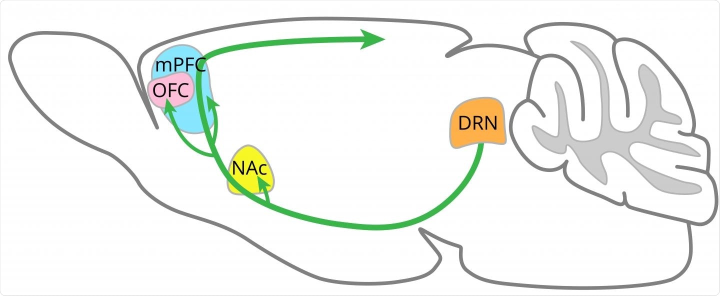
Delayed gratification is vital to human survival. It allows us to suppress our impulses to behave in ways that gain instant gratification to reap rewards later. While it is a vital human cognitive strategy, little is known about how patience is regulated in the brain.

Image Credit: OIST
Previous studies have implicated the role of serotonin but have not uncovered how it interacts with different brain structures to promote patience. Now, a new study conducted at the Okinawa Institute of Science and Technology Graduate University (OIST), Japan, has revealed that the serotonin release in the orbitofrontal cortex and the medial prefrontal cortex is vital in the regulation of patience.
Serotonin’s impact on distinct brain areas
Serotonin has been the focal point of psychological studies for many decades. Much is understood about how the neurotransmitter influences vital regulating human functions such as sleep-wake cycles, appetite, and even moods. It is also implicated in psychological disorders such as depression.
Previous studies have also hinted at its role in regulating patience. Previous rat models have demonstrated that serotonin at least in part controls for impulse-related behavior and that its impact on corticobasal ganglia circuits may be important for this.
Following up from this previous research, the team at OIST designed a study to uncover the neural correlates of patience, and understand how serotonin influences it. The team bred mice who had been genetically engineered to have neurons that release serotonin in response to a light stimulus.
The results of the study, published this month in the journal Science Advances, show that stimulating these neurons resulted in the mice being able to wait longer periods for food, this effect was strongest when the chance of receiving a reward was high and the timing of obtaining said reward was uncertain.
The scientists explored particular regions of the brain that they believed may influence delayed gratification. They implanted optical fibers within the dorsal raphe nucleus, as well as in the nucleus accumbens, the orbitofrontal cortex, or the medial prefrontal cortex.
The role of the orbitofrontal cortex and the medial prefrontal cortex
No impact was found of stimulating the serotonin-releasing neural fibers in the nucleus accumbens, suggesting that it is not involved in regulating patience. When stimulating the serotonin-releasing neural fibers in the orbitofrontal cortex and the medial prefrontal cortex, on the other hand, the researchers observed that the mice were able to wait longer. Suggesting both of these areas are important in regulating patience.
The release of serotonin in the orbitofrontal cortex had a similar effect as the release of serotonin in the dorsal raphe nucleus. Serotonin activation in both of these areas was linked with delayed gratification in situations where the time of the reward was fixed, as well as when it was uncertain, but generating stronger results when it was uncertain.
A slight difference was observed in the medial prefrontal cortex, where researchers saw an increase in patience triggered only when the timing of the reward was varied.
The team’s results demonstrate that each of these areas contributes separately to the overall waiting behavior of mice. To explore this further, a computational model was developed to gain a deeper insight. The model found that the release of serotonin in these brain areas increased the mice’s belief that a reward would be obtained in that particular trial, encouraging them to wait longer.
Improving depression medication
Overall, the study is important in expanding our knowledge of how distinct brain regions and networks are impacted by serotonin and, subsequently, regulate patience. Gaining a more thorough understanding of how serotonin impacts different areas of the brain will be vital to improving particular drugs, such as selective serotonin reuptake inhibitors (SSRIs) which are used to treat depression.