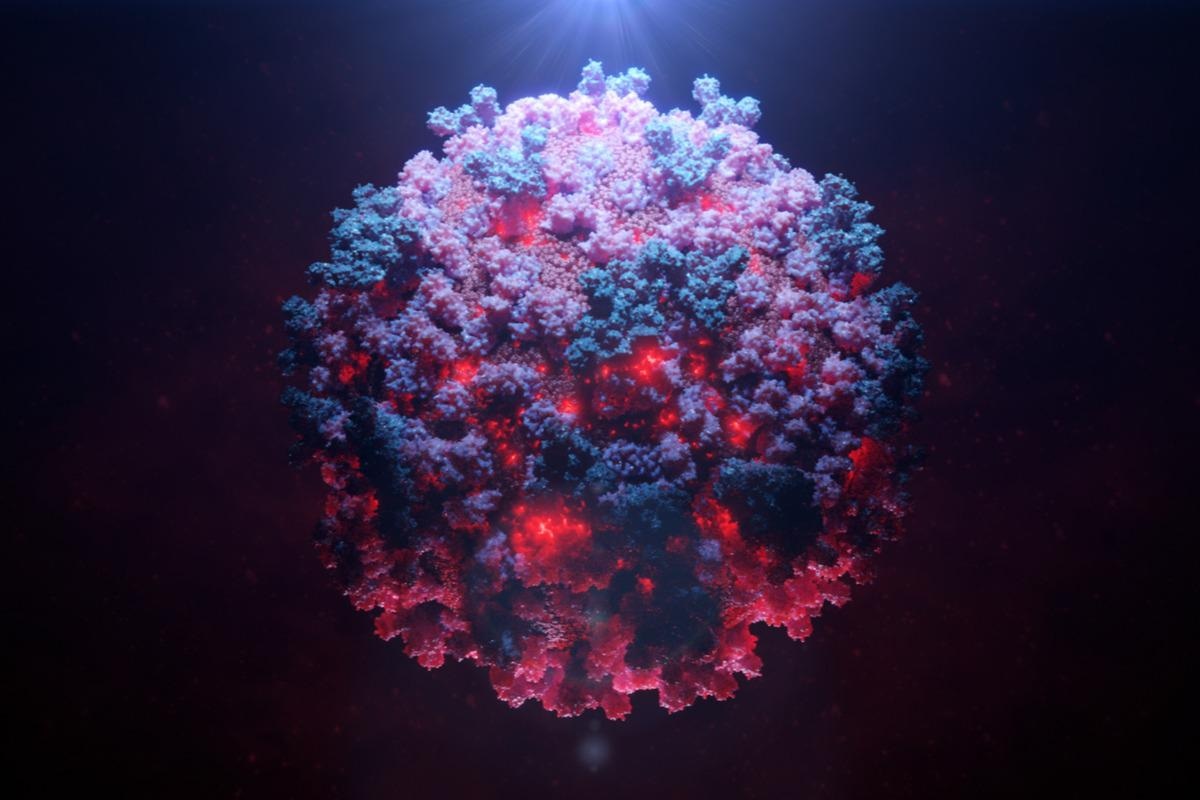
Severe acute respiratory syndrome virus 2 (SARS-CoV-2), the causative agent of coronavirus disease 19 (COVID-19), uses its spike (S) glycoprotein to enter host cells. The S glycoprotein consists of approximately 35% carbohydrate, affecting SARS-CoV-2’s infectivity and susceptibility to antibody inhibition. The S protein is the primary target of neutralizing antibodies elicited by natural infection and by vaccines.
 Study: Glycosylation and disulfide bonding of wild-type SARS-CoV-2 spike glycoprotein. Image Credit: Alpha Tauri 3D Graphics/Shutterstock
Study design
In a recent study posted to the journal Virology, researchers found that virus-like particles (VLPs) produced by co-expression of SARS-CoV-2 S, membrane (M), envelope (E), and nucleocapsid (N) proteins contain S glycoproteins, which are modified by complex carbohydrates. They also studied the disulfide bond and glycosylation profile of S glycoproteins to understand their correct conformation and the composition of the sugar moieties on their surface.
In addition, the researchers evaluated the consequences of natural variations in O-linked sugar addition and the cysteine residues involved in disulfide bond formation. N-linked glycans, such as Asn 234, on wild-type SARS-CoV-2 S glycoprotein trimer are modified in the Golgi-complex of the host cell. The glycans are extracted in pure form using a fucose-selective lectin, a processed glycan, to determine its glycosylation and disulfide bond profile.
Key findings
The native SARS-CoV-2 S glycoprotein was studied in detail using a 293T cell line (293T-S) that expresses the wild-type S glycoprotein under controlled conditions, induced by a tetracycline-inducible promoter. The S glycoprotein was labeled with a carboxy-terminal 2xStrep affinity tag for purification.
Then the 293T-S cells were treated with doxycycline. These cells expressed the S glycoprotein, cleavable into the S1 exterior and S2 transmembrane glycoproteins. These events indicate that the preparation retained the closed conformation of the S glycoprotein. Additionally, they also establish that the purified S glycoproteins bind angiotensin-converting enzyme 2 (ACE2), and nearly all the S glycoproteins on VLPs could be modified by complex carbohydrates.
Mass spectrometry (MS) was used to determine the disulfide bond topology of the purified S glycoproteins. Using this technique, researchers identified disulfide-linked peptides in the tryptic digests of the S glycoprotein preparation. They mapped ten and five disulfide bonds in S1 and S2 and an alternative disulfide bond in S1, between Cys 131 and Cys 136.
The results of mass spectrometry also explained the glycosylation of S glycoproteins in detail. It detected a total of 826 unique N-linked glycopeptides and 17 O-linked glycopeptides. It revealed that O-linked glycosylation occurs at seven glycosylation sites: S659, S673, S680, S1170, T676, T696, and T1160.
The S glycoproteins produced in the GALE/GALK2 293T cell-line formed pseudotype vesicular stomatitis virus (VSV) vectors that exhibited lower infectivity compared to the viruses made by S glycoproteins produced in the 293T cells. Because the post-translational modification of S glycoproteins in the two cell types happens differently, it changes the S glycoprotein function.
The natural variants of SARS-CoV-2 S glycoproteins are rare and are formed by substitutions at one of its cysteine residues, compromising the formation of some disulfide bonds. These variants alter the expression, processing, and functions of S glycoproteins. Upon critically examining these variants and their impact on S glycoproteins, the results indicated that only one Cys15Phe mutant (C15F) remained partially infective, although it was not very stable. The sensitivity of the two most replication-competent S glycoprotein mutants, T676I and S1170F, was analyzed. No significant difference was found in the neutralization sensitivity of the wild-type and mutant viruses.
Conclusions
The study’s results align with previous studies suggesting the predominance of complex carbohydrates of the SARS-CoV-2 S glycoprotein trimer. Compared with the other trimers characterized over time, the wild-type S glycoproteins purified in this study exhibited more glycan processing. The differences observed in glycosylation profiles could be attributed to some specific S glycoprotein constructs used in the study. Nevertheless, the study results enhance the understanding of glycans located on the S glycoprotein trimer by examining their nature and role in the wild-type SARS-CoV-2.
These results give an in-depth knowledge of how these glycans impact natural variation in the glycosylated or disulfide-bonded sites. With enhanced knowledge of SARS-CoV-2 biology and the wild-type S glycoprotein, especially glycans, it would be possible to design better preventive measures such as vaccines and antibody-based therapies against SARS-CoV-2 infections in the future.
Journal reference:
Shijian Zhang, Eden P. Go, Haitao Ding, Saumya Anang, John C. Kappes, Heather Desaire and Joseph G. Sodroski; Glycosylation and disulfide bonding of wild-type SARS-CoV-2 spike glycoprotein. J Virol. doi.org/10.1128/JVI.01626-21 https://journals.asm.org/doi/10.1128/JVI.01626-21