After the 2019 outbreak, severe acute respiratory syndrome coronavirus 2 (SARS-CoV-2) emerged as the largest pandemic since the 1918 influenza outbreak. One of the unique aspects of this virus is that its spike proteins contain a furin cleavage site.
Another novel amino acid motif – QTQTN, is found directly upstream of the FCS, which is also absent in other group 2B CoVs. This motif is often deleted and has been rampant in cultured virus stocks of the alpha, beta and delta variants. Also, the QTQTN deletion was found in a small subset of patient samples.
A new study posted to the bioRxiv* preprint server aimed to demonstrate whether the loss of the QTQTN motif attenuates SARS-CoV-2 replication in respiratory cells in vitro and pathogenesis in hamsters.
 Study: QTQTN motif upstream of the furin-cleavage site plays key role in SARS-CoV-2 infection and pathogenesis. Image Credit: ktsdesign / Shutterstock

 This news article was a review of a preliminary scientific report that had not undergone peer-review at the time of publication. Since its initial publication, the scientific report has now been peer reviewed and accepted for publication in a Scientific Journal. Links to the preliminary and peer-reviewed reports are available in the Sources section at the bottom of this article. View Sources
Findings
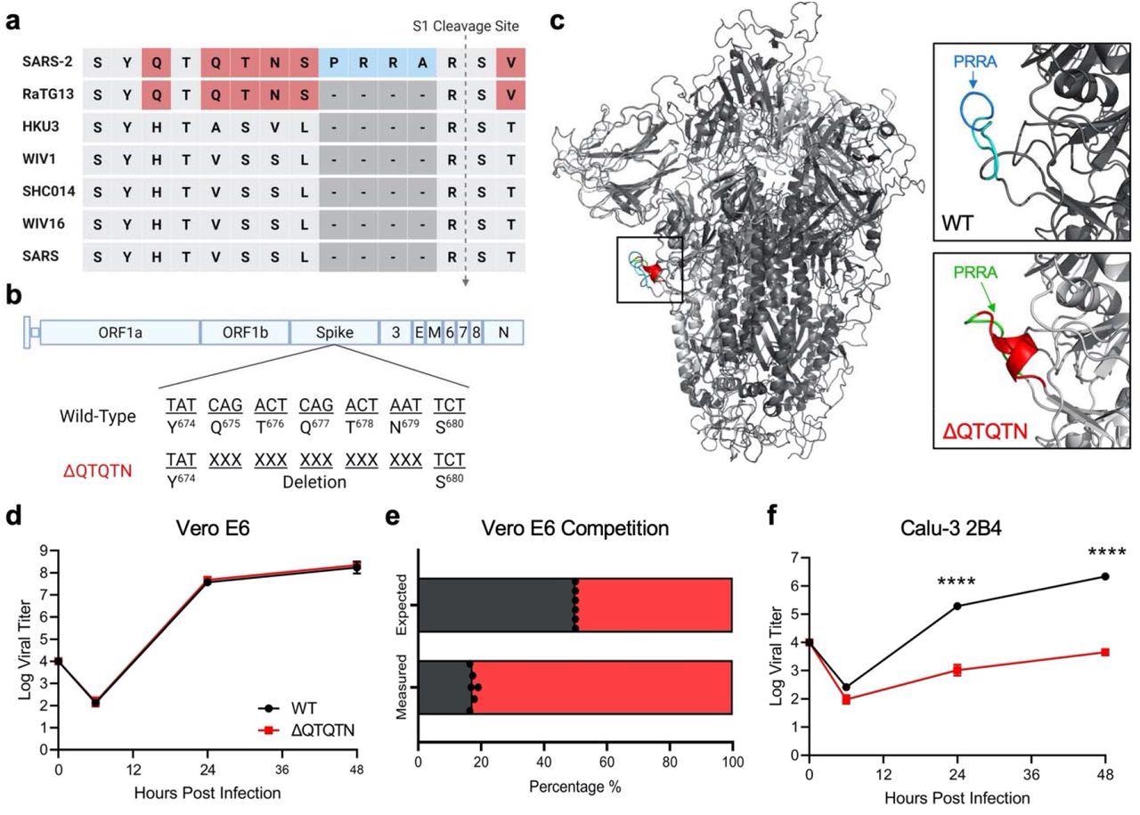
The present experiment entailed a comparison of group 2B coronavirus sequences. It was found that in addition to the furin cleavage site (FCS), a QTQTN motif was present directly upstream in the SARS-CoV-2 spike protein. This motif is absent in other CoVs, except for the closely related RaTG13 69 bat coronavirus. Importantly, this QTQTN motif is often deleted in SARS-CoV-2 strains propagated in Vero E6 cells.
The QTQTN deletion on the SARS-CoV-2 spike structure was examined. The results showed that the ΔQTQTN mutant forms a stable α-helix in the loop containing the S1' cleavage site. This α-helix is speculated to make the loop less flexible and reduce access to the proteolytic cleavage site.
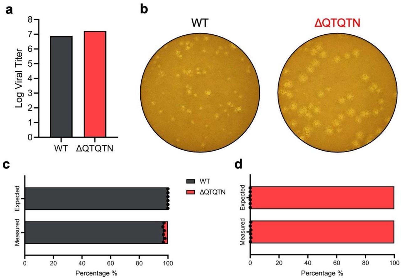
It was also noted that the deletion of the QTQTN motif did not affect virus replication in Vero E6 (African green monkey kidney cells) cells – with the rescue stock titer comparable to wild-type WA-1 (WT) in yield. However, the ΔQTQTN mutant produced a large plaque morphology, as seen with FCS knockout mutant.
 In vitro characterization of SARS-CoV-2 ΔQTQTN. a, Comparison of S1/S2 cleavage site across SARS-CoV, SARS-CoV-2 and 5 related bat CoVs. b, Schematic of SARS-CoV-2 genome with deletion of QTQTN codons. c, SARS-CoV-2 spike trimer (grey) with WT (upper) and model-predicted ΔQTQTN (lower) overlaid. PRRA (blue) is exposed with QTQTN (cyan) present in WT and extends the loop (upper). An α-helix is formed with the deletion of QTQTN (red) and PRRA (green) is exposed (lower). d, Viral titer from Vero E6 cells infected with WT (black) or ΔQTQTN (red) SARS-CoV-2 at an MOI of 0.01 (n=3). e, Competition assay between WT and ΔQTQTN SARS-CoV-2 at a ratio of 1:1, showing RNA percentage from next-generation sequencing. f, Viral titer from Calu-3 2B4 infected with WT or ΔQTQTN SARS-CoV-2 at an MOI of 0.01 (n=3). Data are mean ± s.d. The statistical analysis was measured by a two-tailed Student’s t-test. *, p≤0.05; **, p≤0.01; ***, p≤0.001; ****, p≤0.0001.
On the other hand, the ΔQTQTN mutant had a significant advantage over WT SARS-CoV-2 in Vero E6 cells. This probably caused the accumulation of this mutation in Vero E6-amplified virus stocks.
Notably, Calu-3 2B4 cells – a human respiratory cell line - exhibited a ~2.5 log reduction in ΔQTQTN replication at both 24- and 48-hours post-infection (hpi). Hence, the ΔQTQTN mutant is attenuated in respiratory cells and has a fitness advantage in Vero E6 cells.
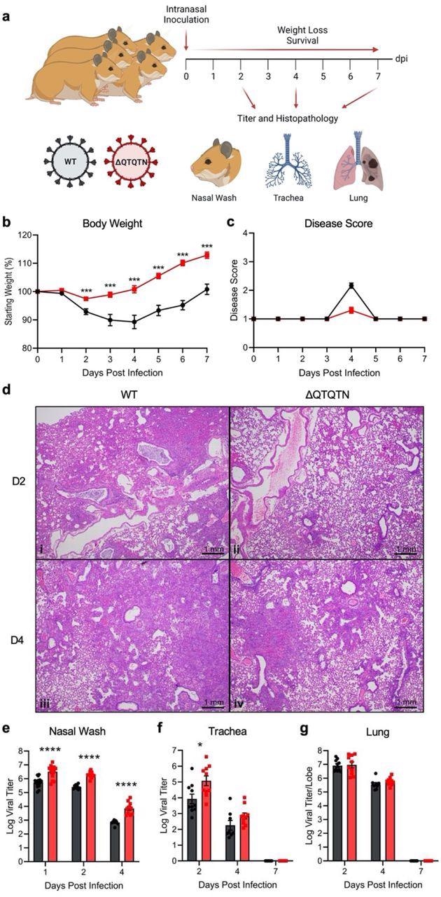
Here, hamsters infected with ΔQTQTN developed less extensive pulmonary lesions than those in hamsters infected with WT SARS CoV-2. All lesions depicted – interstitial pneumonia, peribronchitis, peribronchiolitis, and vasculitis with predominantly subendothelial and perivascular infiltration by lymphocytes and perivascular edema. Meanwhile, cytopathologic effects were observed in alveolar pneumocytes and bronchiolar epithelium. Thus, the deletion of QTQTN motif attenuated coronavirus disease 2019 (COVID-19), in vivo.
 ΔQTQTN SARS-CoV-2 replication. a, Virus stock titer of WT and ΔQTQTN SARS-CoV-2 from Vero E6. b, Plaque morphology of WT and ΔQTQTN in Vero E6. c-d, Competition assay between WT and ΔQTQTN SARS-CoV-2 at a ratio of 100:0 (c) and 0:100 (d) WT:ΔQTQTN, showing RNA percentage from next generation sequencing.
However, ΔQTQTN viral replication in vivo was not compromised compared to WT SARS-CoV-2. In fact, the viral titers were more significant – with a 10-fold increase in nasal wash titers at 1, 2, and 4 dpi. Similar titer elevations were identified in the trachea of infected hamsters. Notably, viral titers were equivalent in the lungs for 2 and 4 dpi.
On examining the viral ribonucleic acid (RNA), the ΔQTQTN had comparable levels of viral replication relative to control SARS-CoV-2 in the lung. While hamster lung samples elicited clustering of WT and ΔQTQTN at 2 dpi and 4 dpi. In addition, upregulated genes were similar between WT and ΔQTQTN compared to mock at both time points. Therefore, the attenuation of ΔQTQTN in vivo is not due to a change in replication capacity – these data are consistent with in vivo results with the FCS knockout virus.
Furthermore, loss of the QTQTN motif resulted in minimal S1/S2 cleavage product and greater full-length spike compared to WT control. Similar suppression of spike processing was seen in Calu3-2B4 cells, which was still higher than in Vero E6. The deletion of the QTQTN motif appeared to impair spike cleavage at the S1/S2 site.
 In vivo characterization of SARS-CoV-2 ΔQTQTN in golden Syrian hamsters. a, Schematic of golden Syrian hamster infection with WT (black) or ΔQTQTN (red) SARS-CoV-2. b-c, Three- to four-week-old male hamsters were infected with 105 plaque-forming units (pfu) of WT or ΔQTQTN SARS-CoV-2 and monitored for weight loss (b) and disease score (c) for seven days (n=10). d, Histopathology of hamster lungs manifested more extensive lesions in animals infected with WT SARS-CoV-2 on day 2 (i) (4X magnification) than in animals infected with ΔQTQTN (ii) (4X). Lesions increased in volume on day 4 with greater proportions of the lungs affected in hamsters infected with WT (iii) (4X) than ΔQTQTN (iv) (4X) on day 4. e-g, Viral titers were measured for nasal washes (e), tracheae (f), and lungs (g). Data are mean ± s.e.m. Statistical analysis measured by two-tailed Student’s t-test. *, p≤0.05; **, p≤0.01; ***, p≤0.001; ****, p≤0.0001. Figures were created with BioRender.com
Additionally, the loss of the QTQTN motif reduced the virus's capacity to use TMPRSS2 (present at the cell surface) for entry. Besides, the loss of glycosylation sites in the QTQTN motif attenuates replication in Calu-3 2B4 cells. While the loss of glycosylated residues did not impact the spike processing of SARS-CoV-2.
Whereas, disruption of both glycosylation residues with the QVQVN mutant titer resulted in attenuation—equivalent to that of ΔQTQTN—suggesting that abolishing both O-linked glycosylation sites disrupted TMPRSS2 utilization. Glycosylation of the QTQTN motif appeared essential for protease interactions with spike and SARS-CoV-2 infection.
In conclusion, the FCS, the length/composition of the exterior loop, as well as glycosylation of the QTQTN motif are necessary for the infection and pathogenesis. Disruption of any of these three elements attenuates SARS-CoV-2.
Moreover, the QTQTN deletion results in reduced spike cleavage and diminished capacity for cell surface entry. Mutations of glycosylation-enabling residues in the QTQTN motif also render replication attenuation despite intact spike processing.
The results isolated elements in the SARS-CoV-2 spike and the furin cleavage site that contribute to increased replication and pathogenesis of the virus.

 This news article was a review of a preliminary scientific report that had not undergone peer-review at the time of publication. Since its initial publication, the scientific report has now been peer reviewed and accepted for publication in a Scientific Journal. Links to the preliminary and peer-reviewed reports are available in the Sources section at the bottom of this article. View Sources
Journal references:
- Preliminary scientific report.
Vu, M., Lokugamage, K., Plante, J., et al. (2021), "QTQTN motif upstream of the furin-cleavage site plays key role in SARS-CoV-2 infection and pathogenesis", bioRxiv* preprint, doi: 10.1101/2021.12.15.472450, https://www.biorxiv.org/content/10.1101/2021.12.15.472450v1
- Peer reviewed and published scientific report.
Vu, Michelle N., Kumari G. Lokugamage, Jessica A. Plante, Dionna Scharton, Aaron O. Bailey, Stephanea Sotcheff, Daniele M. Swetnam, et al. 2022. “QTQTN Motif Upstream of the Furin-Cleavage Site Plays a Key Role in SARS-CoV-2 Infection and Pathogenesis.” Proceedings of the National Academy of Sciences 119 (32). https://doi.org/10.1073/pnas.2205690119. https://www.pnas.org/doi/full/10.1073/pnas.2205690119.