Recently, natural severe acute respiratory syndrome coronavirus 2 (SARS-CoV-2) infections are increasingly being reported in various domesticated and captive wild species, while both in-silico and experimental infections of SARS-CoV-2 have uncovered immunogenicity and pathogenesis of the virus in numerous species. Such infections are considered spillover infections and evidence of large-scale veterinary infections are lacking.
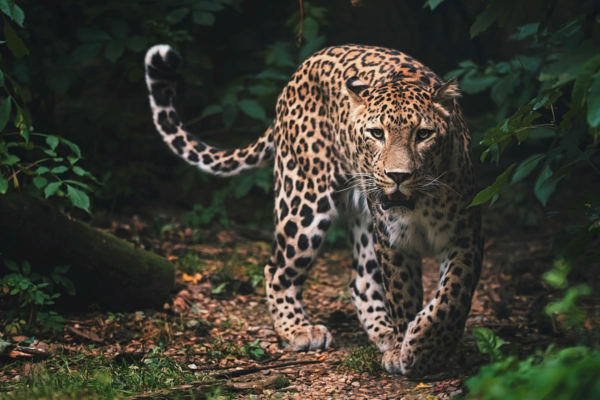
 Study: Systemic infection of SARS-CoV-2 in free ranging Leopard (Panthera pardus fusca) in India. Image Credit: Ondrej Chvatal/Shutterstock
Background

 This news article was a review of a preliminary scientific report that had not undergone peer-review at the time of publication. Since its initial publication, the scientific report has now been peer reviewed and accepted for publication in a Scientific Journal. Links to the preliminary and peer-reviewed reports are available in the Sources section at the bottom of this article. View Sources
Approximately, 160 Km from the national capital of New Delhi, the carcass of about a 1-year-old male leopard cub was recovered from the agricultural field of Mojipur village of Social Forestry, Bijnor range. Necropsy was conducted by ICAR-Indian Veterinary Research Institute following COVID-19 protocol; the examination revealed piercing wounds on both the sides of the ventral neck region (canine teeth marks of prey animal), subcutaneous contusions and hemorrhages on the neck and cranium. In addition, there was a consolidation of both the lungs, along with severe vascular changes like congestion and hemorrhages in the visceral organs.
A nasopharyngeal swab was found positive for SARS-CoV-2 by RT-PCR. The results were confirmed by generating a partial spike protein gene sequence using Sanger’s method.
The positive nasopharyngeal swab was subjected to virus isolation in Vero cells and observed for obvious cytopathic effects (CPE). The presence of the virus was confirmed by real-time polymerase chain reaction (RT-PCR) and immuno-staining with SARS-CoV-2 positive serum, followed by detection by fluorescein isothiocyanate (FITC) labeled anti-human secondary antibodies. Whole-genome sequence was generated directly from the nasal swab specimen through outsourcing to Eurofins and submitted to NCBI.
Spike protein sequence of virus from the leopard had a high resemblance to that of Delta variant and with the sequences generated in a previous study from infected Asiatic lions from Jaipur and Etawah, India. Furthermore, there were two amino acid substitutions (Thr77Lys and Asp142Gly) compared to genome sequences from infected Asiatic lions of Tamil Nadu.
Although very few animals have been detected with SARS-CoV-2 infection, the high resemblance of these veterinary strains with that of human Delta variant suggests possible spillover infection.
Here, representative sequences from the Global initiative on sharing all influenza data (GISAID) database were downloaded from the Global initiative on sharing all influenza data (GISAID) database of each clade of SARS-CoV-2 samples from India. The leopard sample was sequenced and suggested a possible spill over infection with the prevailing Delta mutant. Additionally, the animal’s brain spleen, lymph node, and lung specimens were positive for SARS-CoV-2 with cycle threshold (Ct) values of E-gene—which codes for the structural envelope protein—ranging from 27.5-31.6.
Histopathological analysis exhibited diffuse areas of consolidation, hemorrhages, pneumocyte hyperplasia, septal thickening, and perivascular infiltration of mononuclear cells in the lungs. In addition, there existed severe vascular changes in the heart, brain, liver, and kidneys while the spleen and lymph nodes showed mild depletion of lymphoid follicles.
Immunohistochemistry analysis of the leopard sample disclosed viral antigen in the lungs, brain, and spleen. In addition, septal lining cells, alveolar macrophages, endothelial cells of pulmonary vessels, and bronchiolar epithelial cells also demonstrated abundant antigen. These lung histopathological results were similar to natural infections in minks and humans and experimental infection of SARS-CoV-2.
Conclusions
The findings suggested systemic SARS-CoV-2 infection prior to death and traumatic injuries inflicted by another carnivore were the immediate cause of death. Furthermore, infection of the central nervous system (CNS) was evident by the detection of the virus in the brain section.
Previously, most studies in animals were carried out on animals with a history of human contact, with most of the studies in animals reported either in domestic settings or with captive wild animals with a history of human contact.
Detection of SARS-CoV-2 in a wild leopard emphasizes the need for vigilant screening for the development of carrier status in wild felids.

 This news article was a review of a preliminary scientific report that had not undergone peer-review at the time of publication. Since its initial publication, the scientific report has now been peer reviewed and accepted for publication in a Scientific Journal. Links to the preliminary and peer-reviewed reports are available in the Sources section at the bottom of this article. View Sources
Journal references:
- Preliminary scientific report.
Mahajan, S., Karikalan, M., Chander, V., et al. (2022). Systemic infection of SARS-CoV-2 in free ranging Leopard (Panthera pardus fusca) in India. bioRxiv. doi.org/10.1101/2022.01.11.475327 https://www.biorxiv.org/content/10.1101/2022.01.11.475327v1
- Peer reviewed and published scientific report.
Mahajan, Sonalika, Mathesh Karikalan, Vishal Chander, Abhijit M. Pawde, G. Saikumar, M. Semmaran, P Sree Lakshmi, et al. 2022. “Detection of SARS-CoV-2 in a Free Ranging Leopard (Panthera Pardus Fusca) in India.” European Journal of Wildlife Research 68 (5). https://doi.org/10.1007/s10344-022-01608-4. https://link.springer.com/article/10.1007/s10344-022-01608-4.