The ongoing coronavirus disease 2019 (COVID-19) pandemic, caused by the severe acute respiratory syndrome coronavirus 2 (SARS-CoV-2), has resulted in high mortality and morbidity throughout the world.
Infections from SARS-CoV-2 have been reported to have various clinical presentations from asymptomatic to severe disease and death. However, most SARS-CoV-2 infections are found to result in mild symptoms.
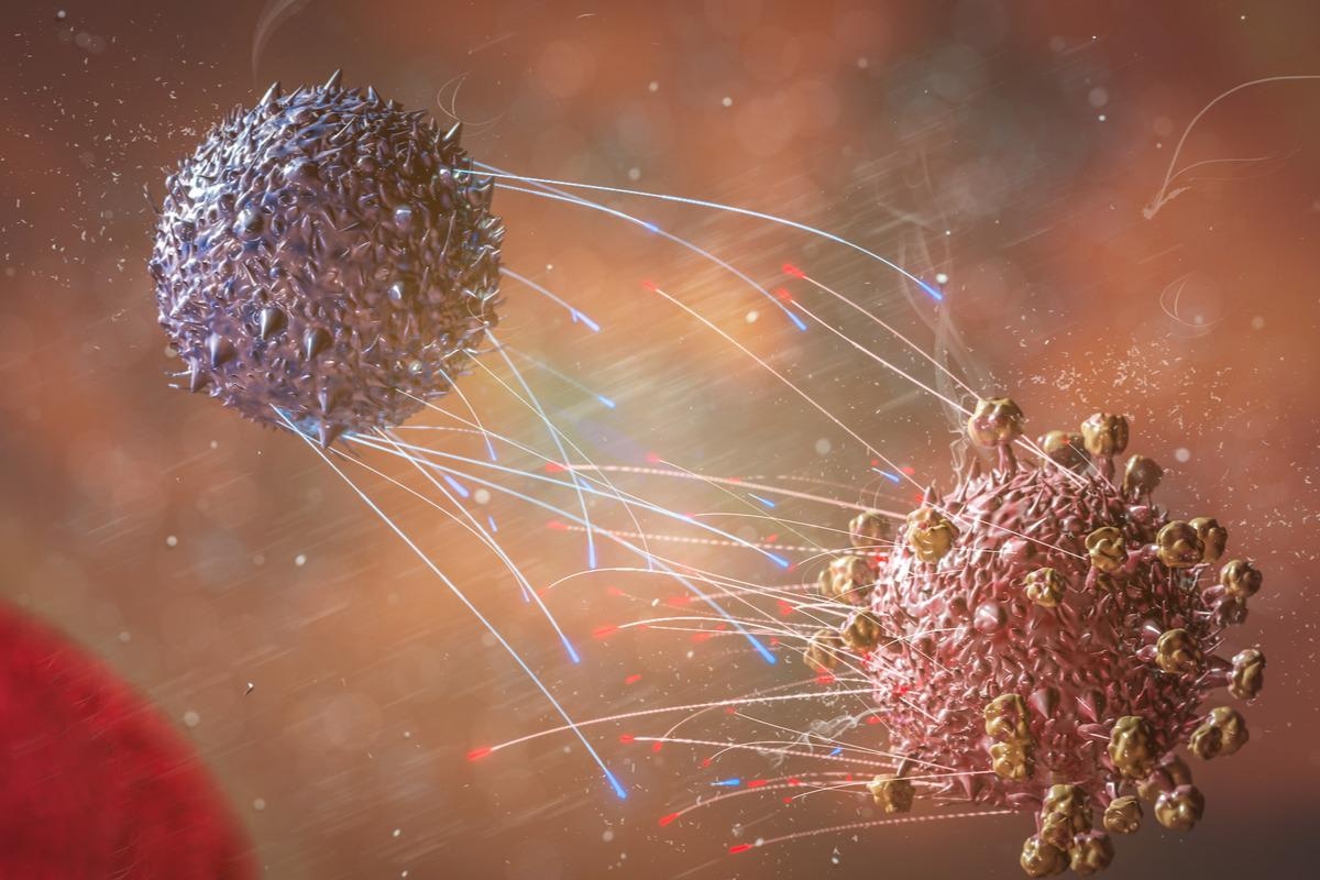
 Study: Broadly-recognized, cross-reactive SARS-CoV-2 CD4 T cell epitopes are highly conserved across human coronaviruses and presented by common HLA alleles. Image Credit: CI Photos/Shutterstock
Older individuals and individuals with comorbidities have been found to develop severe symptoms of disease with high mortality. Genetic polymorphism along with age-related variations in adaptive and innate immunity has been observed to play a critical role in the difference of clinical outcomes.

 This news article was a review of a preliminary scientific report that had not undergone peer-review at the time of publication. Since its initial publication, the scientific report has now been peer reviewed and accepted for publication in a Scientific Journal. Links to the preliminary and peer-reviewed reports are available in the Sources section at the bottom of this article. View Sources
Background
The local and systemic pathogenesis of SARS-CoV-2 can be attributed to reduced type I interferon, deficient anti-viral immunity in nasal epithelial cells, recruitment and activation of neutrophils, and atypical peripheral blood cytokine profile.
The role of T cells is not fully understood yet against this background although studies have shown that low CD4+ and CD8+ T cell counts are associated with severe disease. Peak severity has been observed to be inversely correlated with the frequency of SARS-CoV-2-specific IFN-γ-producing CD8+ T cells. Also, early CD4+ T cell responses were found to be associated with mild disease.
In addition to SARS-CoV-2 that has caused the current pandemic, six other coronaviruses are known to infect humans. These coronaviruses include the highly pathogenic SARS and MERS beta-coronaviruses, less pathogenic alpha coronaviruses 229E and NL63, and beta coronaviruses OC43 and HKU1. Reports after the discovery of the original SARS virus suggest that the T cells from unexposed individuals were capable of recognizing naturally processed and presented SARS antigens even before infection.
Studies have suggested that previous infections with common-cold coronaviruses (HCoVs) may have given rise to cross-reactive SARS-specific responses in unexposed individuals. However, T cells from individuals not previously infected with SARS were found to have an impaired response when compared with CD8+ T cells from previously SARS-infected individuals.
Studies on immune cross-reactivity among the human coronaviruses have gained importance due to the emergence of SARS-CoV-2. Both the SARS-CoV-2-infected individuals and the unexposed individuals were found to possess cross-reactive antibodies to both SARS-CoV-2 and HCoV spike protein. Moreover, individuals who had a recent infection with HCoV were found to have less severe COVID-19 infections.
Several studies suggested that T cell reactivity has been found in up to 50 percent of individuals who have not been exposed to SARS-CoV-2. A recent study reported that several people who had been exposed to SARS-CoV-2 did not test positive for the infection due to early T cell response. Although there is a high prevalence of HCoV infection and sequence homology with SARS-CoV-2, the definite relationship between the pre-infection T cell response and previous HCoV infection is not clear.
A new study published in the pre-print server bioRxiv* investigated SARS-CoV-2 spike protein responses targeted by cross-reactive T cells that were isolated from convalescent COVID-19 individuals along with previously uninfected donors that involved pre-pandemic donors sampled between 2015 to 2018 and seronegative asymptomatic individuals who were sampled during the pandemic.
About the study
The study involved generation of Peptide-pool or individual peptide expanded T cell lines from freshly isolated or frozen peripheral blood mononuclear cells (PBMCs) followed by IFN-γ ELISpot assay. Thereafter, Intracellular cytokine staining (ICS) was performed using the in vitro expanded T cells.
T cell clones were isolated from PBMCs followed by stimulation and blocking assay. A peptide binding assay was carried out to measure spike peptide binding. This was followed by tetramer staining, sorting of DP4-163/164 cells, T-cell receptor (TCR) sequencing, and clonotype analyses.
Study findings
The results indicated that a COVID-19 donor showed strong IFN-γ responses to peptide pools from SARS-CoV-2 spike (S), membrane (M), nucleocapsid (N) but not envelope (E) protein. However, responses to the spike (S) proteins of the four HCoVs were comparatively weaker. Moreover, responses to the SARS-CoV-2 S pool were observed to expand following stimulation with the HCoVs S pool. A pre-pandemic donor also reported IFN-γ T cell responses to S pools from each of the four HCoVs along with SARS-CoV-2 S peptides.
The results of the ex vivo studies were similar to the in vitro studies except for 42 percent of uninfected donors showed positive IFN-γ responses specific for SARS-CoV-2 S pool after in vitro expansion in comparison to 10 percent observed in the case of ex vivo. Ex vivo responses were also observed against HCoVs S pools in most COVID-19 and uninfected donors.
The results also indicated three pairs of overlapping peptides in the SARS-CoV-2 S protein sequence that could serve as epitopes for the cross-reactive T cell responses. These three overlapping peptides were 198/199, 190/191, and 163/164. Among 10 convalescent COVID-19 donors, 5 showed ex-vivo responses to 163/164, and one donor each recognized 190/191 and 198/199. Following in vitro expansion with HCoV S pools, another donor was found positive for peptide 163/164.
Furthermore, the three cross-reactive epitopes were identified from the S2’ domain of the S protein. The 163/164 sequence comprised the S2’ cleavage site and the fusion peptide (FP), 190/191 is located in the first heptapeptide repeat region, and 198/199 is located between heptapeptide repeats 1 and 2. These regions were found to be highly conserved among the SARS-CoV-2 variants including Omicron and Delta.
The T cells that responded to expansion were mostly reported to be CD4+. Therefore, it can be concluded that peptides 163/164, 190/191, and 198/90 contain epitopes presented predominantly by MHC-II proteins. Moreover, both the DP4- restricted clones and DQ-5 restricted clones were found to bind epitopes within the 163/164 sequence with a three-residue register shift between the core epitopes. DQ5-restricted clones were found to recognize 6 to 9 residues long minimal peptide sequences whereas DP4-restricted clones were found to recognize 9 residues long minimal peptide sequences.
Alignment of the DQ5 and DP4 core epitopes showed that the P2, P5, and P8 positions were 100 percent conserved in both SARS-CoV-2 and HCoV sequences. Other residues of DQ5 and DP4 are less conserved which accounts for the binding preference to several homologs. Also, the SARS-CoV-2 163/164 epitope was observed to be recognized broadly in unexposed, convalescent, as well as mRNA-vaccinated donors.
Tetramer staining validated the importance of DP4-163/164 tetramer in the detection of SARS-CoV-2 and HCoV-cross-reactive T cell populations. The results also identified a highly diverse repertoire of TCRs that recognized the 163/164 peptide. The TRAV and TRBV gene sharing among the donors was found to be quite limited.
The current study was capable of identifying a pan-coronavirus epitope that could be responsible for the cross-reactive T cell response. The epitope was found to be highly conserved among the human coronaviruses as well as the SARS-CoV-2 variants of concern. These conserved sequences can be useful in studies that are related to pre-existing HCoV immunity to COVID-19 severity or incidence. Moreover, they must also be considered for inclusion in pan-coronavirus vaccination.
Limitations
The study had certain limitations. First, the non-IFN-γ-secreting populations were not considered in the determination of cross-reactive responses.
Second, in vitro culture conditions may have an impact on the cross-reactive T cell response regarding the involvement of mostly CD4+ T cells.
Third, the sample size of the study was small, and finally, the study did not determine which donors were previously exposed to which HCoV.

 This news article was a review of a preliminary scientific report that had not undergone peer-review at the time of publication. Since its initial publication, the scientific report has now been peer reviewed and accepted for publication in a Scientific Journal. Links to the preliminary and peer-reviewed reports are available in the Sources section at the bottom of this article. View Sources
Journal references:
- Preliminary scientific report.
Artiles, A.B. et. Al (2022). Broadly-recognized, cross-reactive SARS-CoV-2 CD4 T cell epitopes are highly conserved across human coronaviruses and presented by common HLA alleles. bioRxiv. doi: https://doi.org/10.1101/2022.01.20.477107 https://www.biorxiv.org/content/10.1101/2022.01.20.477107v1
- Peer reviewed and published scientific report.
Aniuska Becerra-Artiles, J. Mauricio Calvo-Calle, Marydawn Co, Padma P Nanaware, John Cruz, Grant C Weaver, Liying Lu, et al. 2022. “Broadly Recognized, Cross-Reactive SARS-CoV-2 CD4 T Cell Epitopes Are Highly Conserved across Human Coronaviruses and Presented by Common HLA Alleles.” Cell Reports 39 (11): 110952–52. https://doi.org/10.1016/j.celrep.2022.110952. https://www.cell.com/cell-reports/fulltext/S2211-1247(22)00734-3.