Coronavirus disease 2019 (COVID-19) has spread to nearly every country globally and caused over 5 million deaths. Following the emergence of the initial strain in Wuhan, China, and its sudden international spread, many governments opted to enact costly and restrictive measures to prevent the alarmingly rapid reproduction number of severe acute respiratory syndrome coronavirus 2 (SARS-CoV-2) from rising higher.
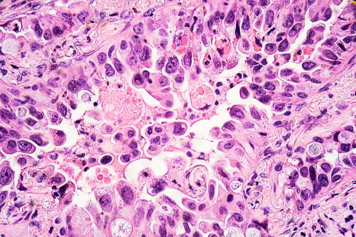
 Study: The effect of COVID-19 mRNA vaccine on respiratory system: human lung carcinoma cells by means of Raman spectroscopy and imaging. Image Credit: David A Litman/Shutterstock
These measures were slowly dismantled once several different vaccines were produced and administered in developed countries. However, threats remain from new variants, and most developing nations do not have vaccinated populations. Researchers from the Lodz University of Technology have been investigating the effects of the mRNA COVID-19 vaccines on human lung carcinoma cells.

 This news article was a review of a preliminary scientific report that had not undergone peer-review at the time of publication. Since its initial publication, the scientific report has now been peer reviewed and accepted for publication in a Scientific Journal. Links to the preliminary and peer-reviewed reports are available in the Sources section at the bottom of this article. View Sources
A preprint version of the study is available on the bioRxiv* server while the article undergoes peer review.
The study
Raman imaging allowed scientists to investigate the cellular components without breaking open the cell. Cytochromes can be classified based on the lowest electronic energy absorption band in their reduced state, allowing the different cytochromes to be distinguished. Laser excitation with a wavelength of 532nm with the energy in coincidence with the electron absorption of the Q band can lead to enhanced intensity of the Raman scattering that allows cytochrome c to be studied at low concentrations under the same physiological conditions as humans.
Cytochrome c is located in the internal mitochondrial membranes between complex III and complex IV, and it is important in the electron transport chain. It normally exists in the oxidized form and is reduced during respiration. Raman spectra are excellent at markers of the redox status of cytochrome c, as the Raman signals of the reduced form are significantly higher than the oxidized form.
The researchers demonstrated that there is no release of cytochrome c to the cytoplasm in lung cancer cells. The number of mitochondria in a single cell decreased upon the incubation with mRNA that determines their metabolic activities. The results of the initial Raman imaging proved that the mRNA vaccine does not block the transfer of electrons between complexes III and IV of the respiratory chain but does decrease the number of mitochondria in cells, which demonstrates lower metabolic requirements and lower efficiency of oxidative phosphorylation and ATP synthesis. A similar effect was observed in brain cancer cells.
The scientists then observed the localization of cytochrome c in the cytoplasm to check if the lower concentration was related to apoptosis. They used Raman imaging to show the effect of the mRNA vaccine on the cytoplasm of human lung carcinoma, which showed the signal for both the control cells and the cells incubated with the COVID-19 mRNA vaccine was the same. This shows that the lower concentration of the cytochrome in the mitochondria is not caused by release into the cytoplasm – which could trigger apoptosis.
The researchers then focused on lipid synthesis in cells incubated with the mRNA vaccine. They showed that the cytochrome c was significantly modified following the incubation. The signals corresponding to C-H bending vibrations of lipids in the rough endoplasmic reticulum and lipid droplets increased significantly. This could indicate that the mRNA alters lipid metabolism following up-regulation by de novo lipid synthesis.
The lipid droplets in normal cells are filled with retinyl esters/retinol and are heavily involved in signaling. The scientists hypothesized that incubation with mRNA increases the role of signaling. The scientists looked for indications that the mRNA could enter the nucleus but, as expected, found nothing. They also found no significant indication of changes in the interactions at the cell surface.
The conclusion
Despite the lack of changes observed in the nucleus of the cell, the authors suggest that the mRNA vaccine changes the cell in ways similar to cancer, adding the caveat that this only affects cytochrome c and lipids. The data presented does suggest an alteration in the behavior of the cells.
Still, the aggressive conclusion could easily be misinterpreted and used as fuel for anti-vaccination beliefs, even though none of the changes observed in these cells could be passed on to daughter cells. As well as this, the levels of vaccine the cells were incubated in were significantly higher than most cells will experience following vaccination. While the conclusion does overreach significantly, the information on the effect of the vaccine on the mitochondria and cellular lipids is valuable and could help inform vaccine manufacturers in the future.

 This news article was a review of a preliminary scientific report that had not undergone peer-review at the time of publication. Since its initial publication, the scientific report has now been peer reviewed and accepted for publication in a Scientific Journal. Links to the preliminary and peer-reviewed reports are available in the Sources section at the bottom of this article. View Sources