In a recent study under consideration at Nature Portfolio Journal and posted to the Research Square* preprint server, researchers demonstrated the evolution of multi-system inflammatory syndrome in children (MIS-C) following acute severe acute respiratory syndrome coronavirus 2 (SARS-CoV-2) infection.
MIS-C is a rare hyperinflammatory immune response to acute SARS-CoV-2 infection observed in children. MIS-C patients exhibit features such as intense cytokine production, particularly interferon-gamma (IFN-γ), and lymphocyte activation, associated with fever and organ dysfunction.
Recent studies have outlined a prominent role for the Notch receptor (Notch) signaling pathways, particularly the Notch4 locus, in association with acute coronavirus disease 2019 (COVID-19) and related respiratory illnesses. It is upregulated on lung tissue regulatory T (T reg) cells in an interleukin (IL)-6-dependent manner and helps subvert tissue repair function in favor of an inflammatory response.
In addition, the Notch family, composed of five ligands (Delta-like 1, 3, 4, Jagged1, and 2), and four Notch receptors (Notch1 to 4) influence conventional T (Tconv) cell responses. However, the underlying immune dysregulation mechanisms governing post-acute COVID-19 syndromes, including MIS-C, remain unknown.
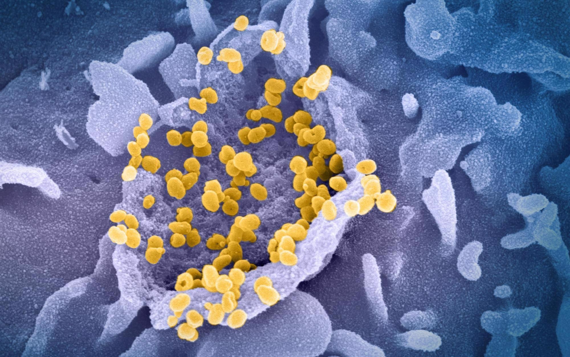
 Study: Notch1-CD22-Dependent Immune Dysregulation in the SARS-CoV2-Associated Multisystem Inflammatory Syndrome in Children. Image Credit: NIAID

 This news article was a review of a preliminary scientific report that had not undergone peer-review at the time of publication. Since its initial publication, the scientific report has now been peer reviewed and accepted for publication in a Scientific Journal. Links to the preliminary and peer-reviewed reports are available in the Sources section at the bottom of this article. View Sources
About the study
In the current study, researchers examined a cohort of 45 children with MIS-C and 50 children with COVID-19 from centers in the United States, Turkey, and Italy. As controls, they evaluated 12 adults with COVID-19, five children with Kawasaki disease (KD), and 18 healthy children.
The team collected the peripheral blood of three MIS-C patients before treatment, another five after treatment, and four healthy controls for single-cell ribonucleic acid (RNA) sequencing (scRNA-seq) analysis to examine the cluster of differentiation 4 (CD4+) T cell dynamics. Further, they used whole-genome sequence (WGS) analysis using Fischer testing and Monte-Carlo simulation for gene enrichment analysis.
All the MIS-C patients met the Centers For Disease Prevention (CDC) case definition and had one or more of the following symptoms- rash, conjunctivitis, and gastrointestinal (GI) symptoms. Moreover, they were highly inflamed, lymphopenic, and coagulopathic. Additionally, over 90% of MIS-C patients showed SARS-CoV-2 seropositivity.
Study findings
The scRNA-seq analysis outlined six subsets of CD4+ T cells; however, performing a graph-based clustering analysis using Seurat revealed 16 more CD4+ T cell clusters.
The authors observed that eight of the 16 clusters (Clusters 1 to 8) were enriched in cells annotated as CD4+ naïve and expressed C-C motif chemokine receptor 7 (CCR7) and selectin L (SELL) genes. Clusters 10 to 14 were enriched in activated CD4+ T cells, such as CD69, with Cluster 10 displaying high nuclear factor kappa B (NF-kB) signaling.
The remaining three clusters encompassed both naïve and activated cells, including one cluster, Cluster 9 defined by viral sensing gene transcripts interferon-Induced Protein With Tetratricopeptide Repeats (IFIT) 2 and IFIT3. Other clusters, Clusters 15 and 16 were enriched in Treg cell transcripts Forkhead Box P3 (FOXP3) and with Mitotic Cells T Cell Receptor Beta Constant 1 (TRBC1).
In mouse models, an immunostimulant, polyinosinic: polycytidylic acid (poly I: C) recapitulated the MIS-C phenotype, especially those actively expressing Notch1 in Treg cells.
Notch1 induction induced the B cell inhibitory receptor siglec 2 (CD22), which destabilized Treg cells and impaired their suppressive function. Finally, the treatment of mice with an anti-CD22 monoclonal antibody (mAb) suppressed the development of systemic inflammation and restored the T reg cells' suppressive function.
These findings indicated how different Notch receptors governing the MIS-C phenotype mobilized T reg cell-specific tissue inflammatory licensing modules. Therefore, interventions targeting the Notch1-CD22 alignment could serve as a therapeutic strategy in MIS-C. Consequently, in patients resistant to standard anti-inflammatory therapies, therapies targeting cytokines involved in Notch1 induction, including anti-CD22 antibodies, might prove effective
Furthermore, the authors found variants in several Notch pathway genes in patients with MIS-C. Importantly, functional mutations in phosphotyrosine binding (PTB) domains of NUMB and NUMBL genes resulted in increased Notch1 expression and signaling. The results of the Monte Carlo simulation and Fischer test validated the role of Notch pathway-related mutations in MIS-C. Exceptionally, MIS-C patients demonstrated an increased CD22 expression on Treg but not Tconv cells, =in mice.
Conclusions
Taken together, the study results could help in the development of a model that traces the evolutionary trajectory of MIS-C. It demonstrated how SARS-CoV-2 infection resulted in an immune dysregulation process that aggravated the systemic inflammation and disrupted tissue-resident T reg cell function.
However, the findings pointed to the reversible nature of this process and ways to reverse it using anti-inflammatory therapies targeting cytokines involved in Notch1 induction, such as CD22.

 This news article was a review of a preliminary scientific report that had not undergone peer-review at the time of publication. Since its initial publication, the scientific report has now been peer reviewed and accepted for publication in a Scientific Journal. Links to the preliminary and peer-reviewed reports are available in the Sources section at the bottom of this article. View Sources
Journal references:
- Preliminary scientific report.
Talal A. Chatila, Mehdi Benamar, Qian Chen et al. Notch1-CD22-Dependent Immune Dysregulation in the SARS-CoV2-Associated Multisystem Inflammatory Syndrome in Children. Research Square preprint 2022, DOI: https://doi.org/10.21203/rs.3.rs-1054453/v1, https://www.researchsquare.com/article/rs-1054453/v1
- Peer reviewed and published scientific report.
Benamar, Mehdi, Qian Chen, Janet Chou, Amélie M. Julé, Rafik Boudra, Paola Contini, Elena Crestani, et al. 2023. “The Notch1/CD22 Signaling Axis Disrupts Treg Function in SARS-CoV-2–Associated Multisystem Inflammatory Syndrome in Children.” The Journal of Clinical Investigation 133 (1). https://doi.org/10.1172/JCI163235. https://www.jci.org/articles/view/163235.