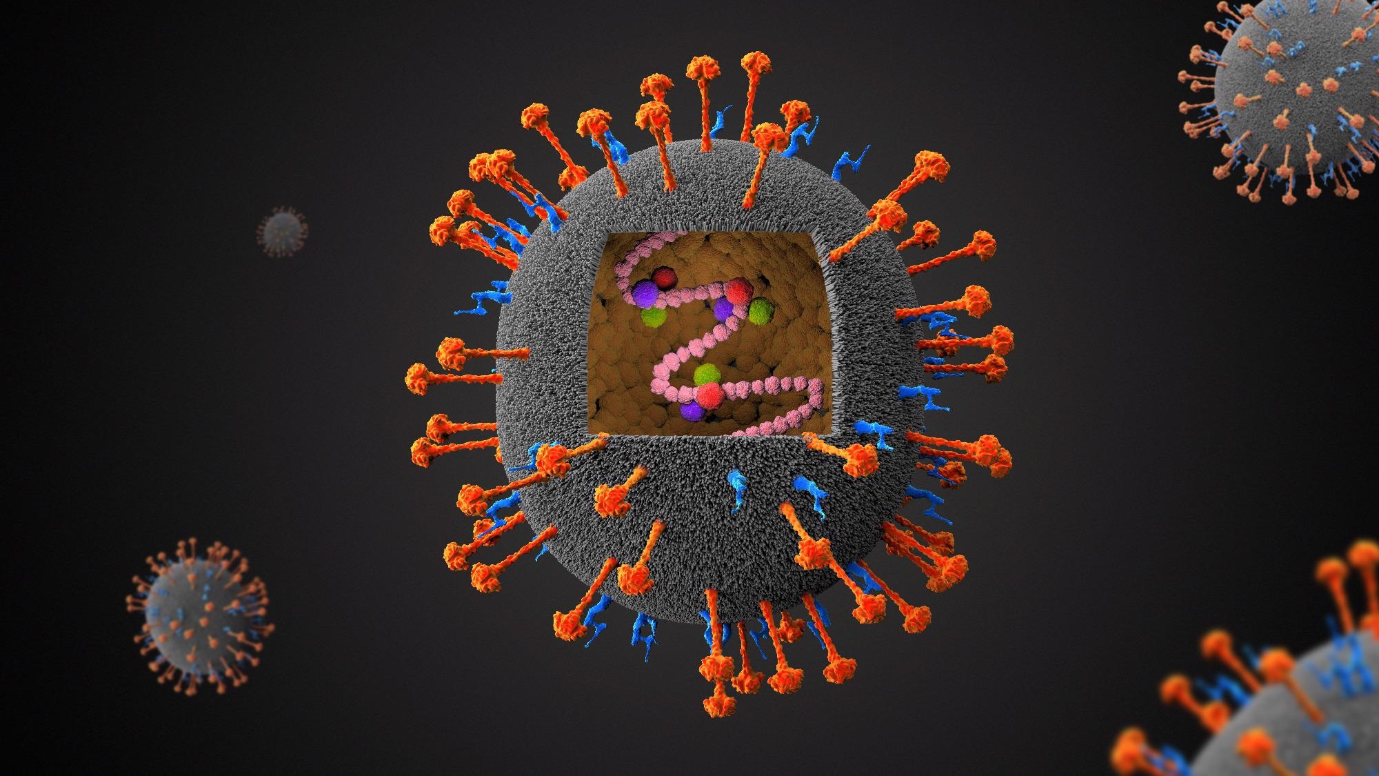
A team of US-based scientists has recently demonstrated the cryo-electron microscopic structure and antigenicity of the attachment glycoprotein of the Nipah virus. The study is published in the journal Science.
 Study: Architecture and antigenicity of the Nipah virus attachment glycoprotein. Image Credit: Festa / Shutterstock
Background
Nipah virus and Hendra virus are zoonotic pathogens belonging to the Henipavirus genus. The viruses cause severe and often fatal respiratory infections and encephalitis in humans, with fatality rates ranging from 50% to 100%. Outbreaks of Nipah virus have been observed frequently in Bangladesh, India, and the Philippines. Recently, a novel Hendra virus genotype has been identified. However, no clinically-approved vaccines and therapeutics are available against Henipaviral infections despite high prevalence and disease severity.
The attachment and fusion glycoproteins on the Henipavirus surface play an essential role in viral entry into host cells. The transmembrane protein tyrosine kinases ephrin-B2 and ephrin-B3 are the host cell receptors responsible for the entry of Henipaviruses. Both attachment and fusion glycoproteins on the viral surface are the primary targets of broadly neutralizing antibodies. In preclinical studies, administration of soluble tetrameric ectodomain of Hendra virus attachment glycoprotein has been found to induce cross-reactive neutralizing antibodies against both Nipah and Hendra viruses.
In the current study, the scientists have derived the cryo-electron microscopic structure of the tetrameric ectodomain of the Nipah virus attachment glycoprotein in complex with a broadly neutralizing antibody. They have also determined the antigenicity of the attachment glycoprotein by vaccinating non-human primates with the tetrameric ectodomain construct.
Structure of tetrameric attachment glycoprotein of Nipah virus
The cryo-electron microscopic structural analysis revealed that the attachment glycoprotein of the Nipah virus forms a 200-Å-long and 120-Å-wide intertwined homo-tetramer. The core of the glycoprotein comprises an N-terminal four-helix bundle (stalk domain) and an interlaced beta-sandwich (neck domain). The C-terminal head domain present on each protomer is connected to the beta-sandwich structure. The entire tetrameric structure of the Nipah virus attachment glycoprotein is stabilized by disulfide bonds formed in the stalk and neck domains.
Further structural analysis revealed that the receptor-binding region of one of the four head domains is oriented towards the host cell surface. In contrast, the other three receptor binding regions are oriented towards the viral membrane. Overall, the tetrameric structure of the Nipah virus attachment glycoprotein was found to adopt a specific conformation with two head domains in the “up” position and two head domains in the “down” position.
Neutralization of Nipah and Hendra viruses
A mouse monoclonal antibody-mediated neutralization of Nipah and Hendra viruses was studied. The mouse antibody showed high efficacy in neutralizing the viruses at nanomolar concentrations, comparable to that observed for a human monoclonal antibody.
The structural analysis of the antigen-antibody complex revealed that the Fab fragment of the antibody binds to the head domain of the attachment glycoprotein, and the heavy and light chains interact with an antigenic region located opposite the receptor-binding region. This indicates that the antibody does not influence the interaction between viral attachment glycoprotein and host receptor. The findings of the membrane fusion assays indicated that the antibody targets the viral membrane – host cell membrane fusion process to inhibit the viral entry into host cells.
The virus neutralization assays conducted using a cocktail of two non-overlapping anti-glycoprotein antibodies revealed synergistic neutralization of Nipah and Hendra viruses. Importantly, the antibody cocktail was found to restrict the emergence of neutralization escape mutations.
Identification of neutralizing epitopes
The purified tetrameric glycoproteins of Nipah and Hendra viruses were used to immunize non-human primates. The serum samples collected from these animals were used to determine the polyclonal antibody response against the viruses. The findings revealed induction of robust neutralizing titers against both viruses at day 84 post-vaccination.
Although full-length tetrameric ectodomain of the glycoprotein was used for immunization, the vaccine-induced antibodies were found to specifically target three distinct epitopes in the head domain of Nipah virus glycoprotein. This indicates that the receptor-binding head domain is the most immunogenic viral component.
Study significance
The study describes the structure and antigenicity of the Nipah virus attachment glycoprotein. These findings are vital for developing next-generation vaccines with improved structural stability and immunogenicity.