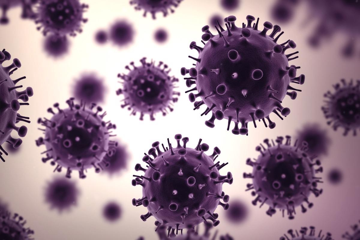
Medical and historical reports suggest that 50 to 100 million people died worldwide during the 1918 influenza A (H1N1) pandemic. The disease was first identified in the summer of 1918 across several continents, peaked during the autumn of 1918, and continued till the winter of 1919. Exceptionally high mortality was reported in 20 to 40 years old healthy individuals, along with severe disease in the elderly and young children.
 Image Credit: Archival influenza virus genomes from Europe reveal genomic variability during the 1918 pandemic. Image Credit: MP Art/Shutterstock
Background
Although it was speculated in 1918 that a virus caused the pandemic, it was confirmed in the 1930s. Studies from the 1990s revealed that the causative agent of the 1918 pandemic was an influenza A virus (IAV) of the H1N1 subtype. Reconstruction of two complete IAV genomes from victims who died in September 1918 at Camp Upton, New York (CU) and in November 1918 at Brevig Mission, Alaska (BM) has taken place since then. Additionally, one complete and 15 partial sequences of the hemagglutinin (HA) gene was obtained from patients who succumbed to the disease between May 1918 and February 1919 in the US and UK.
The studies suggested that at least seven segments were obtained from the diversity of IAV strains that circulated in an avian reservoir and were passed to the seasonal H1N1 influenza viruses. Reconstruction of the 1918 virus indicated that the HA and polymerase complex genes were major determinants of virus pathogenicity. However, several questions regarding the 1918 pandemic remain unanswered.
A new study published in Nature Communications implemented recent advances in nucleic acid recovery to sequence one complete and two partial 1918 influenza virus genomes from specimens sampled in Berlin and Munich.
About the study
The study involved the collection of 11 formalin-fixed lung specimens obtained from the Berlin Museum of Medical History at the Charité, and two were obtained from the Natural History Museum in Vienna, Austria. After that, RNA was extracted from different lung areas, followed by library preparation, sequencing, and NGS data analyses. Finally, evolutionary analyses, simulations, and functional analyses were carried out.
Study findings
The results identified IAV reads in 3 out of the 13 specimens that date to 1918 were associated with histopathological findings of bronchopneumonia. Reconstruction of one complete IAV genome, MU-162 (Munich), and two partial genomes, BE-572 and BE-576 (Berlin), were reported. Several bacteria that have been previously associated with respiratory diseases were also identified in the specimens.
The two partial specimens from Berlin differed at a maximum of two sites in HA. Additionally, the comparison of the nearly complete BE-572 genome with MU-162 identified 22 SNPs. In general, the presence of genomic variability was detected in genomes that were sampled on different continents and at different periods. Furthermore, a two-fold higher activity of BM was observed as compared to MU-162 polymerase. This difference in activity was mostly due to five mutations across the subunits PA (3), PB1 (1), and PB2 (1) that reduced the activity polymerase as compared to BM.
The results suggest that the virus predominantly spreads by local transmission with frequent long-distance dispersal. Also, the presence of amino acid residue G222 in the receptor-binding domain of the H1 subtype HA protein was associated with the increased severity of the virus between the pre-and pandemic peak period. Two other amino acid changes were observed between the pre-and pandemic peak periods, avian-like residues at position 16 (G) and 283 (L) of nucleoprotein were observed for pre-peak strains, while D16 and P283 were observed in peak strains.
Furthermore, seven seasonal H1N1 segments were found to have originated in birds, while one was from a co-circulating homosubtypic H1 IAV. Moreover, the human seasonal H1N1 and 1918 pandemic viruses were found to cluster together with nesting of all internal gene segments of the human seasonal lineage within the pandemic flu diversity.
Conclusion
Therefore, the current study indicates a pure pandemic origin of seasonal H1N1 viruses. However, further research needs to be carried out to ensure this study's findings over the alternative scenario of a homosubtypic reassortment.
The study has certain limitations. First, the sample size of the study is quite small. Second, the pathological specimens are rare and difficult to localize.