In a recent study published in Cell Reports, researchers assessed the impact of severe acute respiratory syndrome coronavirus 2 (SARS-CoV-2) envelope (E) protein on the bromodomain and extraterminal domain (BET) proteins.
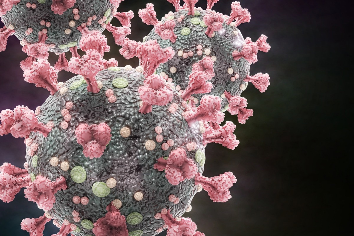
 Study: Viral E Protein Neutralizes BET Protein-Mediated Post-Entry Antagonism of SARS-CoV-2. Image Credit: Dotted Yeti/Shutterstock
Background
The BET family of proteins comprises BRD2, BRD3, BRD4, and BRDT genes. The interaction of the BET proteins with histones as well as cellular transcriptional machinery is known to be essential in several cellular functions such as chromatin remodeling, cell proliferation, and gene expression. Various studies have shown that BRD2 plays an important role as a transcriptional regulator of angiotensin-converting enzyme 2 (ACE2). Thus, using BET inhibitors can decrease ACE2 expression and subsequent SARS-CoV-2 infection
About the study
In the present study, researchers analyzed the function of BET proteins during the course of a SARS-CoV-2 infection.
The post-entry role of the relevant BET proteins was assessed by depleting the proteins A549 cells that overexpressed ACE2 (A549-ACE2). The team developed polyclonal knockout (KO) cells of BRD3 and BED2 as well as knockdown (KD) cells of BRD4 by the nucleofection of clustered regularly interspaced short palindromic repeats (CRISPR) -Cas9 ribonucleoprotein (RNP) complexes. The nucleofection incorporated multiple guide ribonucleic acids (RNAs) against each target. The control cells were incorporated with RNP complexes without an RNA guide. The team verified the deletion of BET proteins via western blotting before the cells were infected with SARS-CoV-2.
The team also generated KO and KD BET proteins in Calu3 cells and airway epithelial cells having ACE2 expression that was sufficient to support viral infection. The KDs and KOs were further validated via western blotting of the BET protein expression, followed by SARS-CoV-2 infection. The impact of depletion of BET proteins on the production of interferon during SARS-CoV-2 infections was tested by infecting Calu3 cells and analyzing the messenger RNA (mRNA) expression of interferon-β (IFNB1), cytokine interleukin 6 (IL6), and interferon-stimulated gene 15 (ISG15). Furthermore, the team utilized the Calu3 BET KD and KO cells to test whether the depletion of BET proteins caused the suppression of gene induction.
The impact of SARS-CoV-2 E protein phenocopies on the BET inactivation was assessed by transfecting an E protein expression construct or an empty control vector (EV) into A549 to stimulate interferon production.
Results
The study results showed that the depletion of BRD3 and BRD4 substantially decreased the viral RNA titers associated with the cells as well as the production of infectious particles in plaque assays. Moreover, only BRD4 KD increased viral replication in cells that were SARS-CoV-2-infected at multiplicities of infection (MOI) of 0.01, while BRD2 KO also increased viral infection at all MOIs to a comparatively lesser magnitude. This suggested that BET proteins played distinct functions in SARS-CoV-2 infections.
The team also observed that the BRD2 KO cells reduced the transcription levels of ACE2 by approximately 80%, which led to a decline in viral RNA levels as well as infectious viral titers. BRD3 KO also decreased ACE expression by almost 50% and viral replication by approximately three-fold. Overall, the team found that BDR4 KD displayed the lowest effect on the expression of viral RNAs but increased the production of infectious particles. This highlighted the impact of BET protein cells on pre- as well as post-entry steps involved in SARS-CoV-2 infection.
Furthermore, the team found robust inductions of the IFNB1, IL6, and ISG15 genes among control cells associated with increasing MOI. On the other hand, the expression of these genes substantially decreased post-treatment with JQ1 or dBET6. Moreover, SARS-CoV-2 infection of the RNP-only control cells displayed a robust stimulation of the three genes at all MOIs. However, the loss of all BET proteins resulted in a remarkable reduction in the expression of the genes at low MOIs. Notably, BET depletion incident at higher MOIs also reduced the induction of IL6 and IFNB1, while ISG15 expression did not show any reduction in cells with a loss of BRD3 and BRD4.
The team also found that the SARS-CoV-2 E protein interacted with BRD2 as well as BRD4. Generation of strep-tagged, acetylation-null point mutants of the E protein, namely K53R, K63R, and K53/63R, showed that the K53/63R mutant reduced binding to BRD2, which suggested that the E protein facilitated the interaction with BRD2.
Conclusion
Overall, the study findings highlighted the antiviral function played by BET proteins throughout the course of a SARS-CoV-2 infection and their distinct roles across different viral life stages. The researchers urge caution regarding the clinical usage of pan-BET inhibitors in the treatment of SARS-CoV-2-infected individuals.