The ongoing coronavirus disease 2019 (COVID-19) pandemic presents challenges in treating infected people and survivors who experience persistent symptoms. Although multiple organs are affected due to severe acute respiratory syndrome coronavirus 2 (SARS-CoV-2) infection, mental health problems are prominent, given that approximately 78% of COVID-19 survivors experience neuropsychiatric long COVID symptoms.
Anxiety, cognitive deficits, and depression are commonly reported, impairing the quality of life. There has been speculation that patients who exhibit severe symptoms during acute infection are more likely to suffer long COVIDs. So far, most neuroimaging studies in COVID-19 patients were performed during the acute stage, and evidence for long-term structural changes in the brain is limited.
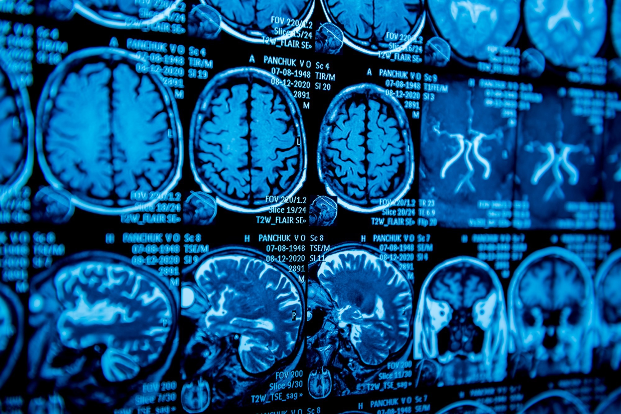
 Study: Larger gray matter volumes in neuropsychiatric long-COVID syndrome. Image Credit: Roman Zaiets / Shutterstock
About the study
In the present study, researchers compared GMVs of patients experiencing neuropsychiatric long COVID symptoms with those of healthy controls. Between April and September 2021, 30 patients with neuropsychiatric long COVID symptoms and 20 healthy controls without a past diagnosis of COVID-19 were included. Patients were recruited from the post-COVID outpatient clinic at the Jena University Hospital.
Inclusion criteria included patients with fatigue, depression, memory, or concentration impairment after COVID. No patient had past psychiatric disorders before COVID-19. Healthy participants were recruited from the community as controls through press releases and tested for anti-SARS-CoV-2 antibodies at the time of scanning.
All participants completed the state-trait-anxiety inventory (STAI), a self-rating assessment for anxiety. In addition, subjects were rated for depression symptoms using Montgomery-Asberg Depression Rating Scale (MADRS). Further, Montreal cognitive assessment (MoCA) was used to screen subjects for cognitive impairment.
The participants underwent a high-resolution T1-weighted magnetic resonance imaging (MRI). The computational anatomy toolbox 12 (CAT12) of the structural brain mapping group, Jena University, was used for voxel-based morphometry. The images were segmented into white matter (WM), gray matter (GM), and cerebrospinal fluid (CSF).
A general linear model (GLM) approach was applied to compare GMVs between patients and controls. Stepwise linear regression was performed to determine if the following variables accounted for the variance in the cortical volumes in patients – gender, age, MoCA, MADRS, STAI, the severity of acute COVID-19, and time since COVID-19 onset.
Findings
Depression and cognitive symptoms significantly differed between long COVID patients and controls at the time of scanning, with a higher burden of symptoms among patients than among controls. Both cohorts had no significant difference in state and trait anxiety. Compared to controls, multiple clusters of larger GMVs in long COVID patients encompassed the hippocampus, insula, frontotemporal regions, amygdala, thalamus, and basal ganglia. In addition, a few clusters of smaller GMVs were present primarily in the bilateral lingual gyri.
The regression model significantly predicted GMVs in patients. The variable, time since COVID-19 onset, significantly predicted GMVs in four clusters. This relationship was inversely correlated, suggesting higher GMVs with a shorter time since COVID-19 onset. Further, gender and age were significant predictors of GMVs in all clusters, implying a linear relationship. MADRS, MoCA, and STAI scores were not significant predictors of GMVs.
Conclusions
The study found increased GM in long COVID patients with neuropsychiatric symptoms. Age, gender, and time since COVID-19 significantly predicted volumetric changes in patients. Larger GMVs in individuals with long COVID might indicate compensatory or recovery effects. Specifically, these effects could be considered for regions connected to the olfactory system, which is believed to be first infected by SARS-CoV before infecting the central nervous system via retrograde neuronal transport mechanisms.
Besides compensatory processes, larger GMVs in patients with COVID-19 could occur from an inflammatory activity resulting in microvascular dysfunction, endothelial activation, and vasogenic increase of tissue water. Because time since COVID-19 onset was a significant inverse predictor of GMVs, this could indicate that larger GMVs may decrease in volume over time. It is also possible that decreasing volumes may be part of COVID-19 recovery.
Overall, the study found that long COVID-19 patients with neuropsychiatric symptoms exhibit changes in GMVs, which are predicted by the individual’s gender, age, and the time since SARS-CoV-2 infection. Further investigations classifying long COVID patients according to the severity of cognitive symptoms and depression could provide better insights into the recovery process.
Journal reference:
- Besteher B, Machnik M, Troll M, et al. Larger gray matter volumes in neuropsychiatric long-COVID syndrome. Psychiatry Research, 2022. DOI: 10.1016/j.psychres.2022.114836