In a recent study posted to Research Square* preprint server, researchers assessed the effect of severe acute respiratory syndrome coronavirus 2 (SARS-CoV-2) and SARS-CoV envelope (E) proteins on human immunodeficiency virus type 1 (HIV-1) infectivity.
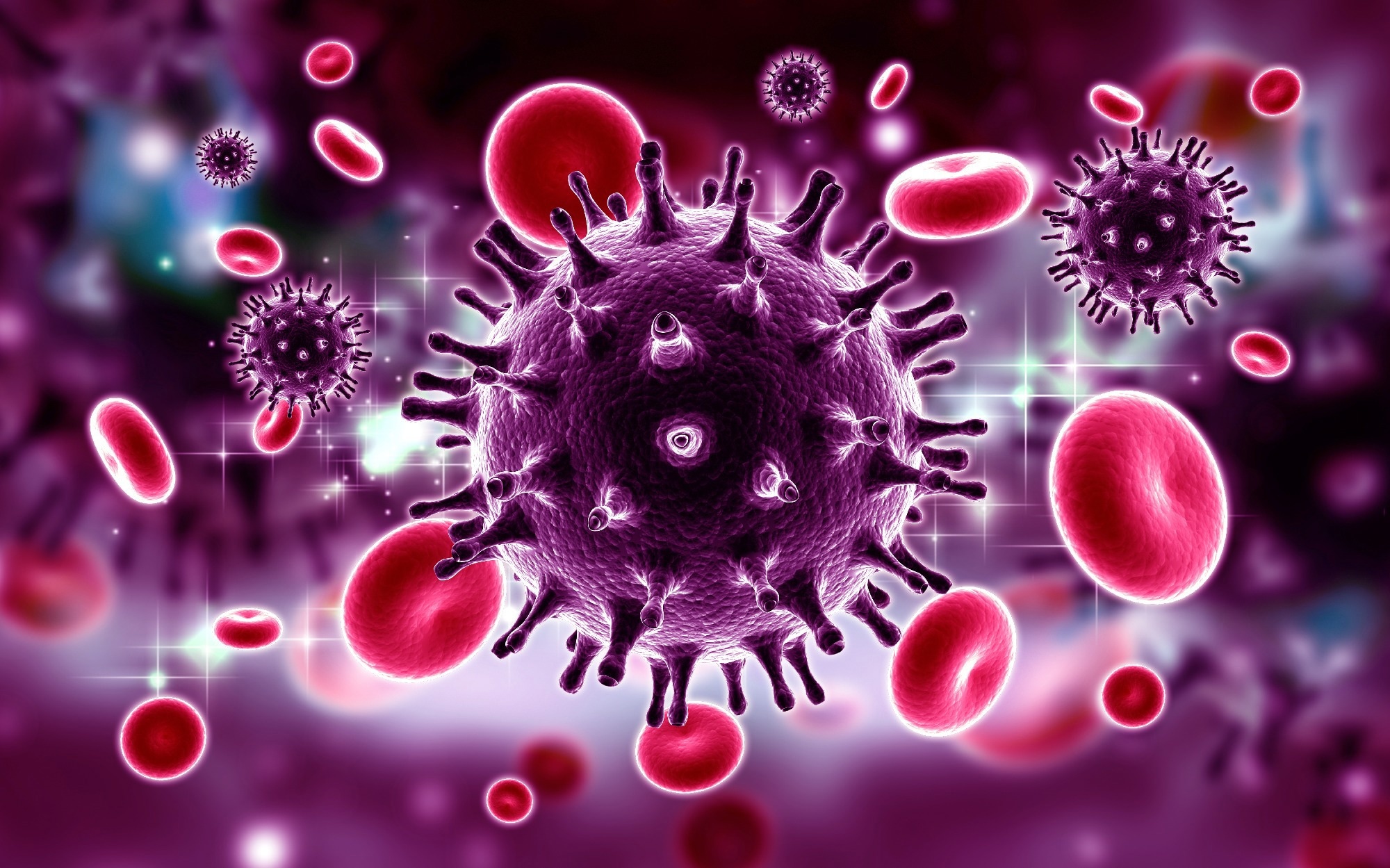
 Study: The Envelope Proteins from SARS-CoV-2 and SARS-CoV Potently Reduce the Infectivity of Human Immunodeficiency Virus type 1 (HIV-1). Image Credit: RAJ CREATIONZS/Shutterstock

 This news article was a review of a preliminary scientific report that had not undergone peer-review at the time of publication. Since its initial publication, the scientific report has now been peer reviewed and accepted for publication in a Scientific Journal. Links to the preliminary and peer-reviewed reports are available in the Sources section at the bottom of this article. View Sources
Ion channels that are virally encoded are called viroporins and are involved in the assembly and release of viruses. The influenza A virus and HIV-1 both encode for viroporins. SARS-CoV-2 encodes for a minimum of two viroporins: the envelope (E) protein and open-reading frame (ORF)-3a. Concerning size, position in the membrane, ion channel activity, and roles to promote virus release, CoV E proteins resemble the viral protein U (Vpu) of HIV-1. Because of these similarities, the team determined the biological characteristics of the SARS-CoV-2 E protein concerning HIV-1 to assess if it could replace the HIV-1 Vpu.
About the study
In the present study, researchers assessed HIV-1 replication in the presence of beta-coronavirus E proteins.
The team first assessed the SARS-CoV-2 E protein's intracellular expression. The SARS-CoV-2 E protein was expressed in COS-7 cells using a vector marked at the C-terminus with a hemagglutinin (HA) tag. The E protein was then immunostained using anti-HA antibodies as well as antibodies against LAMP-1 (late endosomes/lysosomes), endoplasmic reticulum (ER)-Golgi intermediate compartment (ERGIC53), and Golgin97 (trans-Golgi). The team subsequently co-transfected cells with plasmids that expressed E-HA protein and vectors that expressed ER-MoxGFP (RER), TGN38-GFP (TGN), or giantin-GFP (cis/medial Golgi) to study the co-localization of the E protein and the ER, TGN, and cis-medial Golgi. These co-transfections involved fixing cells that were permeabilized and immunostained with an anti-HA antibody to detect E protein.
Additionally, the team compared SARS-CoV-2 E's intracellular localization to SARS-CoV, Middle East respiratory syndrome (MERS)-CoV, and HCoV-OC43 E proteins. The quantity of infectious HIV-1 or vpuHIV-1 produced was then tested to determine whether the SARS-CoV-2 E protein would increase or decrease the quantity. The empty pcDNA3.1(+) vector, which encoded SARS-CoV-2 E protein, herpes simplex virus type 1 (HSV-1) glycoprotein D (gD), which was a positive control for restriction, or gD[TMCT] which was secreted from cells and did not restrict HIV-1, and pNL4-3 were all co-transfected into HEK293 cells.
Results
The study results demonstrated that the ER, ERGIC, TGN, trans-Golgi, and cis-medial Golgi markers co-localized with the E protein. However, neither the cell plasma membrane nor the lysosomes possessed the E protein. This suggested that the three E proteins localized similarly to the SARS-CoV-2 E protein within the cell. The presence of the SARS-CoV-2 E protein dramatically reduced the amount of infectious HIV-1 released. The SARS-CoV-2 E and gD proteins present in the cell lysates were well expressed during the co-transfections, as evidenced by the immunoblots. Furthermore, the team noted that in the absence of the Vpu protein, the protein bone marrow stromal cell antigen 2 (BST-2) decreased the quantity of HIV-1.
The team observed that co-transfection with vectors encoding the vpu HIV-1 genome, BST-2, and the SARS-CoV-2 E protein reduced the infectious virus produced, though not as significantly as the HSV-1 gD protein. Immunoblots of the cell lysates for gD, BST-2, and the SARS-CoV-2 E protein demonstrated that these proteins showed significant expression during the co-transfections. Altogether, the team noted that the SARS-CoV-2 E protein prevented the growth of both viruses and that it could not replace the function of the Vpu protein.
Examining the intracellular expression of these E proteins revealed that, similar to the SARS-CoV-2 E protein, they were exclusively localized in the ER and Golgi sections of the cell without displaying any expression at the cell surface. Although gD was present, it did not affect the amount of infectious HIV-1 released. Instead, it limited its release. The SARS-CoV-2-related and SARS-CoV-related E proteins significantly decreased the amount of infectious HIV-1 released by 1.3% and 1.4%, respectively. A lesser restriction was observed for the MERS-CoV and HCoV-OC43 E proteins at almost 37% and 16% of the pcDNA3.1(+) control, respectively.
The SARS-CoV-2 and MERS-CoV E proteins shared about 37% of their amino acid sequences, while the SARS-CoV-2 and HCoV-OC43 E proteins shared about 26%. The expression of the gD and E proteins was verified by immunoprecipitation from cell lysates obtained from the restriction tests. These findings offer more information about the specificity of HIV-1 restriction.
Overall, the study findings demonstrated the ability of SARS-CoV-2 and SARS-CoV E proteins to impede the development of infectious HIV-1 infection significantly. The study found that protein synthesis was most likely inhibited, and apoptosis was triggered due to SARS-CoV-2 E protein expression. These findings provided evidence that a viroporin from one virus can prevent another virus from infecting cells.

 This news article was a review of a preliminary scientific report that had not undergone peer-review at the time of publication. Since its initial publication, the scientific report has now been peer reviewed and accepted for publication in a Scientific Journal. Links to the preliminary and peer-reviewed reports are available in the Sources section at the bottom of this article. View Sources