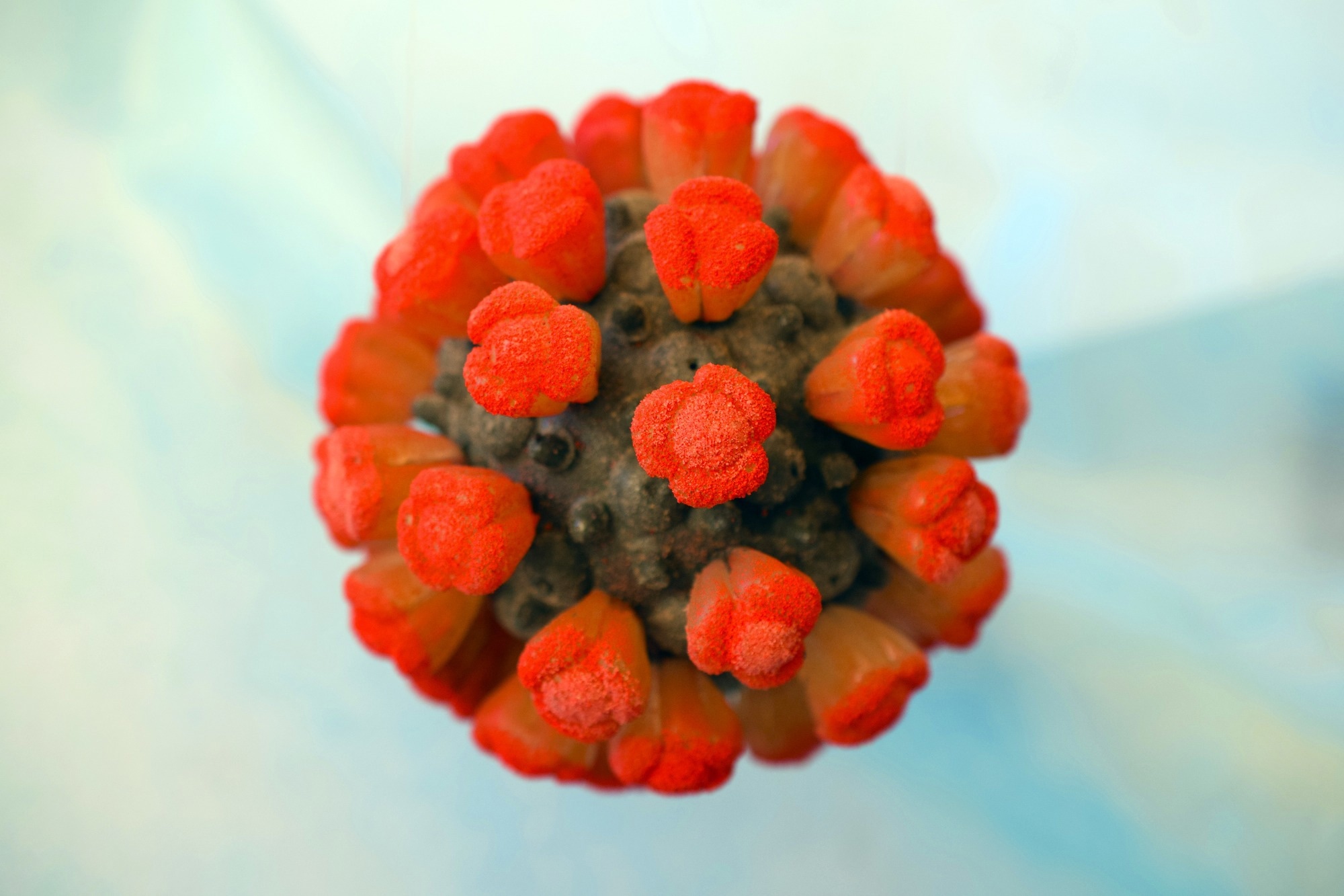
In a recent study posted to the bioRxiv* preprint server, researchers used cellular cryo-electron tomography (cryo-ET) to demonstrate the role of severe acute respiratory syndrome coronavirus 2 (SARS-CoV-2) spike (S) protein in coronavirus disease 2019 (COVID-19)-triggered severe coagulopathies.
 Study: Direct Cryo-ET observation of platelet deformation induced by SARS-CoV-2 Spike protein. Image Credit: Sergejs Pintis/Shutterstock

 This news article was a review of a preliminary scientific report that had not undergone peer-review at the time of publication. Since its initial publication, the scientific report has now been peer reviewed and accepted for publication in a Scientific Journal. Links to the preliminary and peer-reviewed reports are available in the Sources section at the bottom of this article. View Sources
Background
Thrombocytopenia, i.e., low platelet count, myocardial infarction, and non-vessel thrombotic complications, are all commonly observed in COVID-19 patients. In many cases, platelets isolated from COVID-19 patients also show abnormalities, such as hyperactivity and increased spreading behavior.
A recent study suggested the involvement of thrombin and tissue factors (TFs) in the hyperactivation of platelets. When mixed with the SARS-CoV-2 S protein, platelets isolated from healthy donors showed a faster thrombin-dependent clot retraction. In some cases, platelets are activated independently of thrombin with upregulation of signaling factors.
Many theories have reasoned the causes of these abnormalities. Yet, it is unclear whether SARS-CoV-2 directly interacts with platelets, or cytokines, antiphospholipid antibodies, and other immune cells mediate SARS-CoV-2 effects.
Moreover, it is still under debate if platelets have angiotensin-converting enzyme 2 (ACE2) on their surface. Overall, all ultrastructural and patient studies connecting the SARS-CoV-2 severity to the impact of its S protein on platelets have produced fragmented information. It remains unknown what causes the coagulation of platelets in the presence of SARS-CoV-2.
About the study
In the present study, researchers assessed the direct effect of the SARS-CoV-2 S protein on platelet morphology using live imaging and cryo-ET under a collagen type I background. They isolated 119 platelets from healthy de-identified human blood donors. The team performed a solid-phase sandwich enzyme-linked immunosorbent assay (ELISA) and Western blotting to test the role of S protein in platelet activation. In the ELISA assay, they measured platelet factor 4 (PF4) levels, which marked the secretion of alpha-granules, a biomarker of platelet activation. The team employed subtomogram averaging approaches to further analyze the S protein densities on the platelet membrane surface. They manually selected, extracted, and explored the densities from eight tomograms.
Study findings
Discoid-shaped platelets were predominant in the absence of S protein; however, deformed platelets appeared elongated in their presence. Besides extensive elongation, the researchers also noted an increased spreading of platelets in the presence of S protein. They found a population of abnormally shaped platelets resembling a proplatelet-like appearance, indicating an impact on the cytoskeleton-dependent platelet maturation process. Platelets occasionally adhered to extracellular matrix proteins (ECMs) or poly-L-lysine, such as fibronectin and collagen I, without S protein. The researchers hypothesized that integrin receptors mediate SARS-CoV-2 S binding. These receptors on platelets govern filopodia formation crucial for platelet activation. Also, the SARS-CoV-2 S protein contains an arginine-glycine-aspartic acid (RGD) sequence in the receptor-binding domain (RBD), recognized by a subtype of integrin.
The integrin inhibition experiment using cilengitide and in vitro solid-phase binding assays supported this hypothesis, raising the possibility that the S protein recognized integrin αvβ3. However, the observed S-integrin binding was lower than their physiological ligands. Moreover, they did not detect S binding to a platelet integrin, αIIbβ3. Cryo-ET imaging showed actin-rich filopodia at the end of elongated platelets. Unassumingly, there was a dense decoration of S protein on the filopodia surface. An orientation analysis revealed that the S protein bound the platelet membrane surface at various angular distributions.
Furthermore, based on the correlation of S protein binding and filopodia formation, the team assessed the interactions between platelet surface receptors and S protein in vitro. They found a weak but direct interaction of platelet residing RGD ligand integrin receptors αvβ3 and α5β1 with S protein but not with integrin αIIbβ3. Together, these results shed light on the abnormal behavior of platelets leading to coagulopathic events and micro-thrombosis caused by SARS-CoV-2 infection. Several procoagulant players are active during SARS-CoV-2 infection and create a hypercoagulative environment. For instance, the formation of neutrophil extracellular traps, tissue factor 23 (TF23) release, elevation in fibrinogen levels, and dysregulated release of cytokines.
Conclusions
According to the authors, this is the first study directly visualizing the binding of S protein to the platelet surface. The deformation of platelets did not alter their intracellular signaling or induced activation completely. Instead, platelets exposed to S protein needed further stimuli, such as the attachment to an adhesion surface, to get primed for activation. Based on this observation, the researchers speculated that the direct binding of S protein to platelets and other coagulation factors might be responsible for their synergistic and irreversible activation leading to blood coagulation.

 This news article was a review of a preliminary scientific report that had not undergone peer-review at the time of publication. Since its initial publication, the scientific report has now been peer reviewed and accepted for publication in a Scientific Journal. Links to the preliminary and peer-reviewed reports are available in the Sources section at the bottom of this article. View Sources
Journal references:
- Preliminary scientific report.
Christopher Cyrus Kuhn, Nirakar Basnet, Satish Bodakuntla, Pelayo Alvarez- Brecht, Scott Nichols, Antonio Martinez-Sanchez, Lorenzo Agostini, Young-Min Soh, Junichi Takagi, Christian Biertumpfel, Naoko Mizuno. (2022). Direct Cryo-ET observation of platelet deformation induced by SARS-CoV-2 Spike protein. bioRxiv. doi: https://doi.org/10.1101/2022.11.22.517574 https://www.biorxiv.org/content/10.1101/2022.11.22.517574v1
- Peer reviewed and published scientific report.
Kuhn, Christopher Cyrus, Nirakar Basnet, Satish Bodakuntla, Pelayo Alvarez-Brecht, Scott Nichols, Antonio Martinez-Sanchez, Lorenzo Agostini, et al. 2023. “Direct Cryo-et Observation of Platelet Deformation Induced by SARS-CoV-2 Spike Protein.” Nature Communications 14 (1): 620. https://doi.org/10.1038/s41467-023-36279-5. https://www.nature.com/articles/s41467-023-36279-5.