Several patient-derived models of cancer (PDMC), such as patient-derived organoids (PDOs) and patient-derived xenografts (PDXs), have been developed to assess the clinical efficacy of new cancer therapies. The success of precision oncology depends on these models, as they capture molecular, morphological, and functional characteristics of a cancer patient’s tumor, which enables accurate prediction of drug response and resistance.
PDMC can recapitulate antitumor responses in the clinic and, as a result, can provide important insights into personalized care plans.

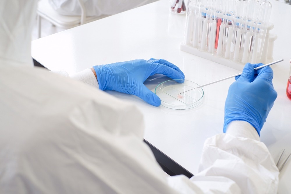
Study: Patient-derived micro-organospheres enable clinical precision oncology. Image Credit: Thananan Kannim / Shutterstock.com
Background
The application of PDMC in clinical decision-making processes is challenging, partly because of the technical limitations within each model. For example, patient-derived cell lines undergo genetic and morphological changes over time, thus making them inappropriate for clinical screening.
Instead, PDXs could be used to analyze intratumoral clonal architecture and genetic diversity, which could help evaluate therapeutic responses. However, this approach has a high operational cost and is slow to generate data, thereby limiting its applicability in diagnostic drug screenings and precision medicine development.
In contrast, PDOs constitute a less costly and higher throughput set of models for clinical applications. In addition, the availability of large-scale biobanks of pancreatic, colorectal, breast, ovarian, kidney, brain, head and neck, and gastric cancers has enabled PDOs to capture patient diversity.
Due to broad-ranging drug screenings, clinicians have detected the associations between genetic mutations and sensitivity to targeted therapies. Therefore, this model has been used as a potential functional precision medicine technology for making appropriate treatment decisions. However, a slow operational process makes it inefficient for clinical use.
Clinical treatment decisions are typically made within 14 days of diagnosis; however, the timeframe for PDO generation exceeds 14 days, which could unacceptably delay treatment. Thus, some of the essential features that must be included in a new diagnostic assay are accelerated PDO generation, functional testing, and automated procedures from the core biopsy.
About the study
The clinical importance of immuno-oncology (IO) has led scientists to focus on reproducing physiological immune activity in organoid cultures. A recent Cell Stem Cell journal study reports the development of an automatic microfluidics droplet platform to rapidly generate PDMC in a high-throughput manner.
The main working principle behind this development involved adding suspended cells from primary tissues to a three-dimensional (3D) extracellular matrix. Subsequently, the cells were mixed with a biphasic liquid (oil) to generate microfluidic-based droplet micro-organospheres (MOS). Finally, the newly generated MOS were demulsified to remove excess oil and were subsequently cultured as suspension droplets.
Key features of the benchtop machine used to generate MOS included reservoirs to load oil and samples directly onto a custom microfluidic chip. Subsequently, the sample outlet was positioned on the backside of the chip for direct dispensing into a MOS recovery vessel.
The attached pressure sources controlled the flow of the oil and sample into the custom microfluidic chip. At the “T” junction of the device, the sample and oil met, wherein the sample was converted into droplets by the oil phase that ultimately reached the collection chamber.
This newly developed device supports a wide range of temperatures, including between the 4 °C sample and 37 °C collection blocks. The chamber height of the device is designed to allow for the generation of MOS of an average of 250-450 mm in diameter. These dimensions are appropriate for several cell numbers and sizes.
An important aspect of the device is its ability to generate MOS of around 15,000 cells from an extremely small sample size of 18-gauge core biopsies. Notably, this sample size is unsuitable for the effective generation of conventional organoids for therapeutic purposes within the constrained clinical timeline.
Study findings
For proof of concept, this device was used to generate MOS from colorectal cancer (CRC) PDX cells. The growth of CRC MOS was monitored from different seeding densities, which revealed the establishment of tumorsphere-like structures by MOS.
The size and number of tumorspheres increased with the seeding density per droplet. Interestingly, MOS generated from clinical CRC biopsies exhibited various morphologies.
Patient-derived MOS were based on microfluid-based technology capable of recapitulating the tumor microenvironment. The MOS were developed from minimal patient tissue to provide reliable results within seven to 14 days of obtaining a biopsy.
Conclusions
The current proof of concept CRC clinical study included only eight patients. As a result, this pilot study provided initial data that supports the potential use of MOS in therapeutics. Although the MOS-based precision oncology pipeline could determine tumor drug responses within 14 days, a large-scale study is required to validate these findings further.
A key strength of this technique is the generation of thousands of MOS from a small biopsy tissue sample. However, more clinical studies must be conducted to validate the clinical value of MOS in terms of specificity of drug response, sensitivity, and reproducibility.
Journal reference:
- Ding, S., Hsu, C., Wang, Z., et al. (2022) Patient-derived micro-organospheres enable clinical precision oncology. Cell Stem Cell 29(6);905-917. doi:10.1016/j.stem.2022.04.006.