In a recent study posted to the Research Square* preprint server, researchers assessed the functional and microstructural brain abnormalities, tiredness, and cognitive impairment after mild coronavirus disease 2019 (COVID-19).
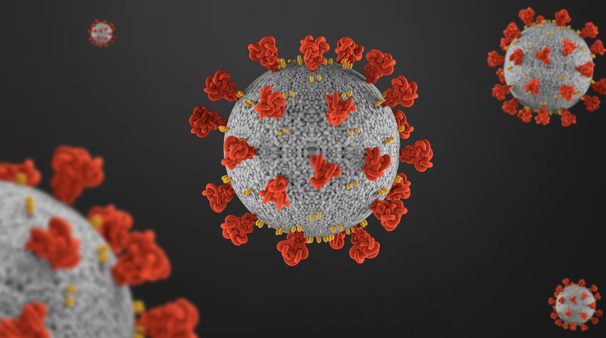
 Study: Functional and microstructural brain abnormalities, fatigue, and cognitive dysfunction after mild COVID-19. Image Credit: GEMINI PRO STUDIO/Shutterstock

 *Important notice: Research Square publishes preliminary scientific reports that are not peer-reviewed and, therefore, should not be regarded as conclusive, guide clinical practice/health-related behavior, or treated as established information.
Studies have identified neurological symptoms of COVID-19, but little is known regarding the long-term neurological effects of severe acute respiratory syndrome (SARS-CoV-2) infection. While most people recover after respiratory symptoms, the prognosis for post-COVID-19 exhaustion and cognitive dysfunction is questionable.
Although neuroinvasion after SARS-CoV-2 has been confirmed in some brain autopsies, the neuronal processes generating neuropsychiatric and neurological symptoms remain unknown.
About the study
In the present study, researchers revealed functional connectivity abnormalities, clinical evaluation, and neuropsychological assessments.
The team performed a cross-sectional data analysis from an observational study aimed at examining post-acute neurological signs and neuroimaging changes associated with COVID-19. utilized social media to publicize its online survey. The first 87 participants who did not need hospitalization and had a confirmed COVID-19 diagnosis were required to visit the center and complete the protocol's four steps: a personal interview and neurological assessment, a 3T magnetic resonance imaging (MRI) acquisition along with neuropsychological assessment, and collection of blood sample at the University of Campinas Hospital. COVID-19 was diagnosed using polymerase chain reaction (PCR) or verified immunoglobulin (Ig)-M or IgG antibodies. A total of 55 healthy volunteers did not exhibit symptoms of COVID-19 and had never tested SARS-CoV-2-positive.
The team conducted an exploratory neuropsychological assessment on recovered patients. Tests were chosen to examine distinct cognitive areas, including language with the Phonemic Verbal Fluency Test and the Verbal Categorical Fluency Test, episodic memory with the Logical Memory subtest of the Wechsler Memory Scale-Revised, and cognitive flexibility with the Trail Making Test.
The team determined the z-scores corresponding to the neuropsychological test results using the Brazilian standard and scaled scores. Supplementary analysis was also performed with multiple linear regression residuals to adjust for the impact of age or education. For each test, the function was classified as preserved, low-average, below-average, or severely low. We measured anxiety and depression symptoms using the Beck Anxiety Inventory (BAI) and Beck Depression Inventory-II, respectively (BDI-II). The Chalder Exhaustion Questionnaire (CFQ-11) and the Epworth Drowsiness Scale were also utilized to assess fatigue as well as excessive daytime sleepiness (ESS), respectively.
Results
The study findings showed a median latency of 54 days between diagnosis and personal interview for the 87 patients we investigated. Patients had an average of four symptoms during the acute phase, while they had an average of two symptoms at the post-acute phase. Fatigue, headache, memory problems, anosmia, and drowsiness were the most common post-acute effects. Moreover, 38 participants felt exhaustion, frequently accompanied by other symptoms like a headache in 25 respondents, memory problems in 17 subjects, and drowsiness in 14 subjects.
The team discovered anomalies in 11 individuals based on post-COVID-19 neurological testing and interview. Along with personal interviews, 65 respondents scored a median of 15 points on the CFQ-11 and 9 points on the ESS. In contrast to the number of symptoms noted at the interview, symptoms indicated exhaustion in 44 of 65 persons and excessive daytime sleepiness among 23 of 65 individuals.
The neuropsychological estimation was conducted on a subgroup of 78 people. Almost 18% of individuals displayed symptoms of depression, while 29% displayed symptoms of anxiety. The team discovered a relationship between BDI-II and CFQ-11 scores. Concerning cognitive function, the team observed abnormal phonological fluency performance among 33% of patients, abnormal trail-making test (TMT)-A performance in 30% of patients, and abnormal TMT-B performance in 40% of patients. An association was also observed between greater fractional anisotropy (FA) values in patients and lower axial diffusivity (AD), mean diffusivity (MD), and radial diffusivity (RD) values.
Conclusion
The study findings showed that SARS-CoV-2 impacted the brains of patients who did not need hospitalization, with chronic fatigue, headache, memory impairments, and somnolence even two months post-SARS-CoV-2 infection. The team identified these patients' cognitive impairment, mild white matter, and connectivity abnormalities. The brain changes and severity of cognitive impairment necessitate substantial longitudinal research on persistent neuropsychiatric symptoms in COVID-19-recovered patients, even in those with minimal acute symptoms. After the initial phase, specific symptom therapy and neurorehabilitation procedures may be required to enhance the cognitive function and quality of life of patients with permanent disabilities.

 *Important notice: Research Square publishes preliminary scientific reports that are not peer-reviewed and, therefore, should not be regarded as conclusive, guide clinical practice/health-related behavior, or treated as established information.