In a recent study posted to the bioRxiv* preprint server, researchers examined the pathological impacts of the structural components of severe acute respiratory syndrome coronavirus 2 (SARS-CoV-2) on human periodontal cells and tissue to understand the association between coronavirus disease 2019 (COVID-19) and deteriorating oral health.
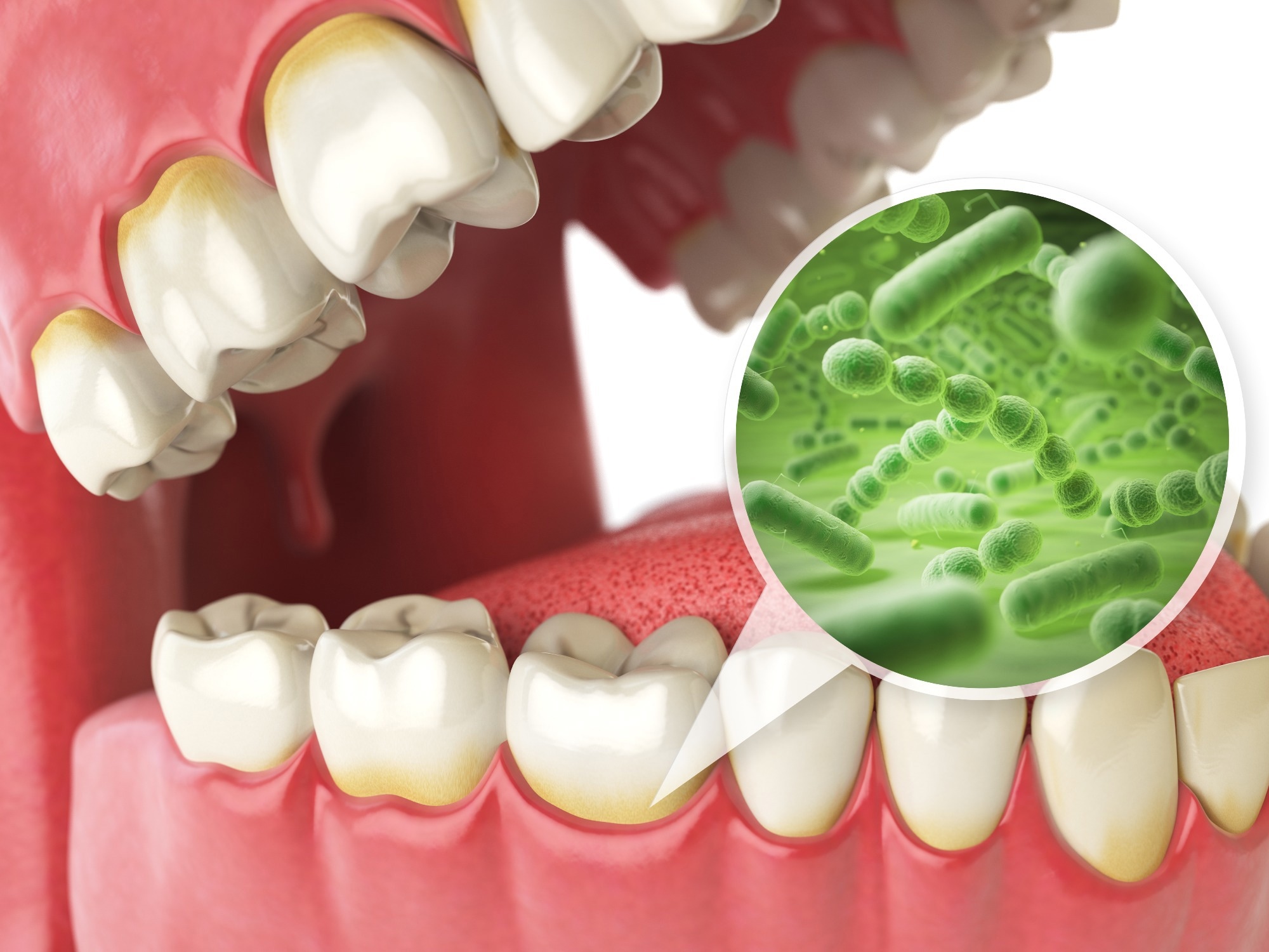
 Study: SARS-CoV-2 causes periodontal fibrosis by deregulating mitochondrial β-oxidation. Image Credit: Maxx-Studio/Shutterstock

 *Important notice: bioRxiv publishes preliminary scientific reports that are not peer-reviewed and, therefore, should not be regarded as conclusive, guide clinical practice/health-related behavior, or treated as established information.
Background
Recent studies have shown that the symptoms of long coronavirus disease (COVID) manifest beyond the pulmonary system, with persistent complications also occurring in the cardiac, renal, and neurological systems. Along with nasal swabs, saliva samples have been used to test for SARS-CoV-2, and the oral cavity is thought to be a viral reservoir. COVID-19 could pose a serious risk to periodontal health since periodontal tissues are vulnerable to infectious diseases. However, the pathology of deteriorating periodontal health due to COVID-19 remains unclear.
The spike protein region of SARS-CoV-2 is the most studied structural part of the virus since most mutations that give rise to variants with increased transmissibility, and immune evasion occur in the receptor binding domain of the spike protein. The spike protein has also been the major target of vaccines and monoclonal antibody therapies. However, SARS-CoV-2 also consists of three other structural components — the envelope, membrane, and nucleocapsid proteins — whose pathology roles have not been comprehensively explored.
About the study
In the present study, the researchers used cultured human periodontal ligament fibroblasts (HPLFs) and human gingival epithelial cells (HGEPp) to investigate the pathology of COVID-19-related periodontal fibrosis.
Immunofluorescence analysis was used to detect the expression of angiotensin-converting enzyme-2 (ACE-2) and transmembrane serine protease 2 (TMPRSS2) receptors in gingival epithelial and periodontal ligament cells, which was confirmed using Western blot analysis. The potential of SARS-CoV-2 to infect HPLFs was explored by treating the cells with SARS-CoV-2 spike protein conjugated with a polyhistidine tag (His Tag). Anti-His Tag allophycocyanin (APC) conjugated antibodies were then used to locate the spike protein through immunofluorescence analysis.
To investigate whether COVID-19 caused fibrosis in periodontal tissues, HPLFs were either infected with lentiviruses carrying plasmids for the envelope, membrane, and nucleocapsid proteins of SARS-CoV-2 or treated with recombinant SARS-CoV-2 spike proteins. Acute and long infections were stimulated in the periodontal fibroblasts, and the cell proliferation was evaluated through immunostaining for anti-Bromodeoxyuridine (BrdU) antibodies. Additionally, Western blotting was used to determine the production of collagen I and metalloproteinase-1 (MMP1) in the extracellular matrix to assess periodontal ligament tissue integrity.
The periodontal ligament fibroblasts treated with SARS-CoV-2 structural components were subjected to proteomic analysis to determine molecular mechanisms regulating COVID-19 pathology. Additionally, the Seahorse Mito stress test was conducted to understand the effects of SARS-CoV-2 infections on the function of mitochondrial fatty acid pathways. The results were validated using etomoxir, which inhibits the mitochondrial β-oxidation pathway.
Results
The results revealed that ACE-2 and TMPRSS2 were expressed in high levels in the periodontal tissues, and exposure to SARS-CoV-2 structural components such as the membrane and envelope proteins increased hyperproliferation of periodontal fibroblasts, along with apoptosis and senescence.
The molecular mechanisms mediating the fibrosis of periodontal tissues mainly consisted of the downregulation of β-oxidation in the mitochondria by SARS-CoV-2 membrane and envelope proteins. Other studies have revealed associations between downregulated mitochondrial β-oxidation and fibrosis in lungs and kidneys. The results also highlighted the role of SARS-CoV-2 structural components other than the spike protein in disease pathology. The authors discussed various studies that identified the alternate mechanisms through which the envelope proteins increased the pathogenicity of SARS-CoV-2, including increased pH within the Golgi apparatus and the formation of cation channels.
The researchers also conducted terminal deoxynucleotidyl transferase deoxyuridine triphosphate (dUTP) nick end labeling (TUNEL) to understand the involvement of SARS-CoV-2 structural components in increasing apoptosis and senescence, and the results indicated that the spike or nucleocapsid proteins did not contribute to an increase in apoptosis and senescence, and only the envelope and membrane proteins did. The membrane and envelope proteins also upregulated collagen I production and decreased MMP1 enzyme production in the extracellular matrix, contributing to periodontal fibrosis.
Conclusions
Overall, the study provided novel insights into the impact of SARS-CoV-2 infections on periodontal health. The results revealed that the SARS-CoV-2 membrane and envelope proteins alter fatty acid degradation pathways in the mitochondria that are essential to maintain energy homeostasis. The dysregulation of the mitochondrial β-oxidation pathway leads to the hyperproliferation of periodontal fibroblasts, causing fibrosis, and increased apoptosis and senescence. These results could contribute to a better understanding of the long COVID symptoms manifesting in other organ systems and provide targets for treatment.

 *Important notice: bioRxiv publishes preliminary scientific reports that are not peer-reviewed and, therefore, should not be regarded as conclusive, guide clinical practice/health-related behavior, or treated as established information.