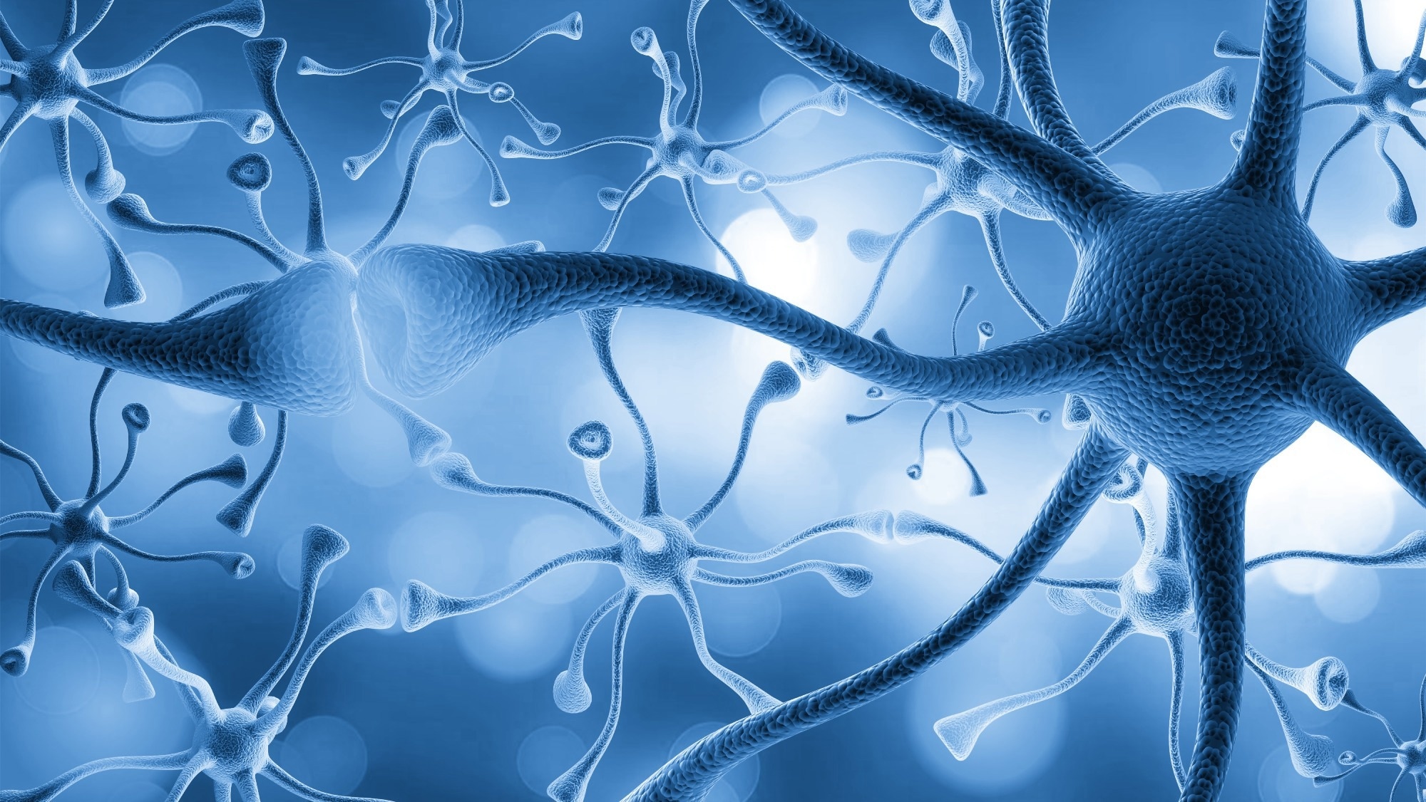
Social interactions need understanding and awareness of others’ behavior. Studies have proposed mirror neurons, or cells denoting actions by self and others, as critical components for cognitive substrates facilitating understanding and awareness. Primate neocortical mirror neurons are reportedly involved in skilled motor tasks; however, the importance of the neurons for their actions and social interactions is unclear. Recent studies have reported that VMHvlPR neurons induce aggression in a social context-sensitive manner, indicating that the moniker attacking center is simple and elides important properties of the neurons.
 Study: Hypothalamic neurons that mirror aggression. Image Credit: Billion Photos / Shutterstock
About the study
In the present study, researchers investigated whether VMHvlPR neurons could perceive aggressive interactions among mice.
A TRAP2-based activity-tagging approach was used to gain genetic access to aggression-mirroring neurons (mirror-TRAP) and investigate their functional relevance in aggressive behavior. VMHvlPR neuronal response among mice engaged in fight was assessed and compared to that among mice who were witnessing aggression.
GCaMP6s were expressed in VMHvlPR neurons of PRCre mice, and fiber photometry was used to imaging VMHvlPR neuronal activity among mice after introducing male intruders in the cage. VMHvlPR neuronal activity among individually hosed males who observed but did not demonstrate aggressive behavior (demonstrators) was also assessed. Aggression mirroring by BNSTTac1 neurons was investigated by expressing GCaMP6s in Tac1Cre male cells and imaging the VMHvlPR neuronal interactions among Trpc2-/- demonstrator mice.
To investigate the need for visual input during aggression mirroring, a perforated partition arena was illuminated using infrared light, to which the murine animals were insensitive. Subsequently, the perforated partition was replaced by a solid, transparent partition to decrease demonstrator mice’s pheromone access to observer mice. Further, mini-scope calcium imaging was performed to investigate VMHvlPR neuronal activity during the enactment and observation of aggression.
GCaMP6s signals of individual neurons were shuttled during the behavioral testing periods to investigate whether the VMHvlPR neurons showed alternate activation and inhibition during the attack and tail-rattling periods. Neuronal co-activation among aggressor and observer mice during the aggressive displays was investigated.
VMHvlPR neurons, whose activity was limited to observers or aggressors during tail-rattles and attacks, were assessed, and the functional importance of observer VMHvlPR cells in territorial fighting was explored. The virally coded, Cre-based inhibitory chemogenetic actuator, DREADDi, was delivered to VMHvl cells, neurons activated among observer TRAP2 male mice were captured, and the males were tested following clozapine-N-oxide (CNO) administration using resident mice-intruder mice territorial fighting assays.
Results
Individual VMHvlPR neurons were activated simultaneously during the observation and performance of attacks and tail-rattles. Visual input, but not prior social experience, was essential for aggression-mirroring by VMHvlPR neurons. The activity of VMHvlPR neurons mirrored aggression between other individuals. Aggression-mirroring VMHvlPR neurons played a specific and essential role in the display of male territorial fighting.
CNO-administered males demonstrated enhanced aggressivity to their mirrors, with three-fold increases in tail-rattling probability and 10-fold increases in tail-rattle numbers. The male mice did not attack mirrors, indicating that other sensory or context cues were needed to induce attacks. The activation of aggressive behavior-mirroring VMHvl neurons intensified male territorial aggression and supplanted male sexual behavior with aggression. The neurons drove aggression toward female, male, and mirror mice.
Neurons whose activity mirrors fighting by other mice were identified in the male mouse hypothalamus and were functionally important for aggression. Mirror neuron activity was found to be critical in fighting, and the forced activation of the neurons triggered aggressive displays by murine animals, even toward their mirror images. The mirror-TRAPed VMHvlPR aggression-mirroring neurons were essential and adequate to elicit male aggression displays. Activation of VMHvlPR neurons was observed during the attacking and tail-rattling forms of aggressive displays.
VMHvlPR neurons exhibited mirroring properties for aggression and such mirroring was not a feature of all populations regulating aggression. The neuronal cells could represent distinct aggressive motor activity and showed largely congruent-type mirroring. The aggression mirror-TRAP strategy effectively captured aggression-mirroring VMHvlPR neurons and could be used to express transgenes of choice in VMHvlPR mirror neurons.
Trpc2-/- VMHvlPR neuronal response during the chemoinvestigation experiments was smaller than their wild-type counterparts, indicating that Trpc2 activity was essential for pheromone signal transduction. The team observed activation of observer VMHvlPR neuronal cells at levels comparable to those across a perforated partition and detected 152 neurons and 88 neurons showing GCaMP6s fluorescence among aggressor mice and observer mice, respectively. The majority (61% to 78%) of VMHvlPR cells were activated among aggressors and observers during bouts of attacks and tail-rattling.
Overall, the study findings showed that VMHvlPR neurons, rather than merely constituting an attacking center, embody a percept of aggression that can trigger fighting in appropriate contexts and monitor third-party aggressive interactions. The findings highlighted the discovery of a non-cortical mirror neuron system in the nucleus of Cajal, a deeply conserved and evolutionary ancient vertebrate hypothalamic region that functionally represents a social behavior and provides a subcortical cognitive substrate critical for social interactions and survival.