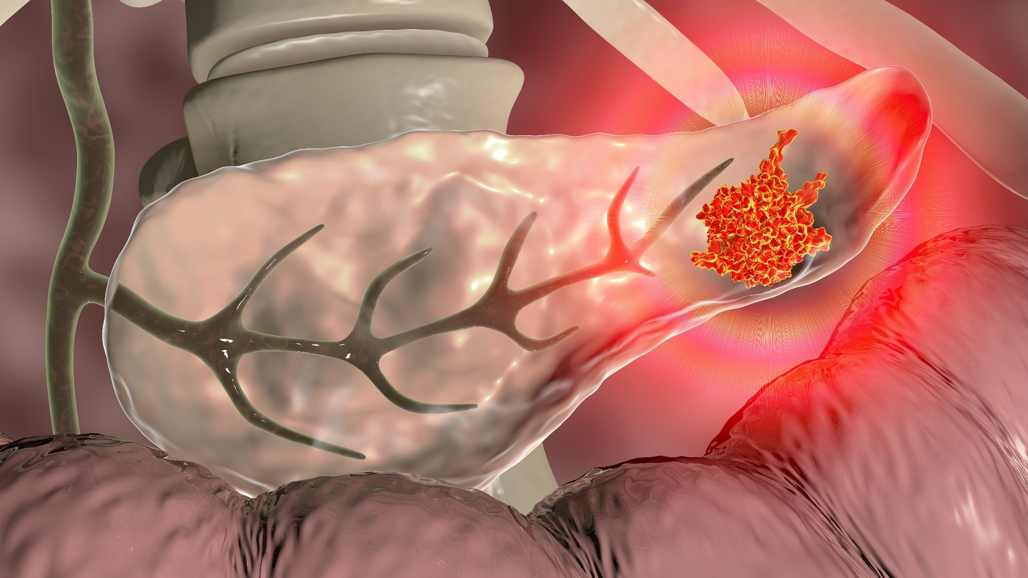
The PDA tumor microenvironment (TME) is characterized by abundant immune cell infiltration, stromal fibroblast expansion, and extracellular matrix deposition. This results in increased interstitial fluid pressure and the collapse of capillaries and arterioles, which collectively contribute to poor oxygen saturation, metabolic aberrations, heterogeneity at the cellular level, and therapeutic resistance. PDA cells in such an extreme TME adapt to increase their catabolic and scavenging capabilities.
 Study: Uridine-derived ribose fuels glucose-restricted pancreatic cancer. Image Credit: Kateryna Kon / Shutterstock
The study and findings
In the present study, researchers screened how metabolites impact the activity of pancreatic cell lines in nutrient-restricted conditions. First, they used a phenotypic screening platform on two non-malignant pancreatic cell lines and 19 PDA cell lines to examine the ability of cells to metabolize over 175 nutrients under nutrient-restricted conditions.
This analysis revealed the utilization of several metabolites under glucose deprivation at comparable levels as the positive control. Most cell lines utilized uridine, adenosine, and other sugars. Next, the team assessed the correlation between metabolite utilization patterns and the expression of associated genes.
Uridine phosphorylase 1 (UPP1) expression correlated with uridine metabolism. This metabolite-gene pair was further studied because uridine was utilized by all PDA cells, and this has not been explored in the context of PDA. The correlation was further validated through mRNA and protein analyses. The utilization of adenosine and inosine did not correlate with UPP1 expression.
Further, the PDA cell lines were sorted into high or low uridine metabolizers. UPP1 was the leading gene in high-uridine metabolizers. Endocytosis and several immune-related or inflammatory pathways were upregulated in high-uridine metabolizers. Moreover, UPP1-high PDA tumors showed elevated expression of glycolysis genes.
UPP1-high PDA cell lines and tumors exhibited downregulation of metabolic pathways, such as glutathione, fatty acid, and amino acid metabolism. Next, the researchers investigated the activity of equimolar uridine and glucose. Uridine and glucose fueled metabolism similarly across four PDA cell lines.
They also observed an increased cellular reducing potential upon uridine supplementation and speculated that uridine-derived ribose might be recycled to support the reducing potential. As such, ribose was supplemented to glucose-deprived cells. Like uridine, supplementing ribose fueled the reducing potential.
The investigators supplemented a UPP1-low or -high cell line with uridine under a glucose-deprived state and applied a metabolomic analysis based on liquid chromatography-mass spectrometry (LC-MS). They found higher glycolytic intermediates, lactate secretion, tricarboxylic acid (TCA) cycle, uridine, and amino acid derivatives in UPP1-high and UPP1-low cell lines.
Further, the team followed the metabolic fate of isotopically-labeled uridine using LC-MS. They noted the labeling of uridine diphosphate (UDP) or triphosphate (UTP), adenine monophosphate (AMP), diphosphate (ADP) or triphosphate (ATP), nicotine adenine dinucleotide (NAD+), non-essential amino acids (serine, glutamate, and aspartate), oxidized glutathione, and the intermediates of glycolysis, TCA cycle, and hexosamine biosynthesis.
Next, the researchers knocked out UPP1 in a UPP1-low or -high cell line and observed that the ability of uridine to rescue cellular bioenergetics was abolished in the absence of glucose. Knocking out UPP1 broadly altered the cellular metabolome in both cell lines. Uridine-derived ribose recycling was also blocked in the knocked-out cells.
The researchers observed an increased expression of UPP1 in public PDA datasets compared to non-tumor samples. Notably, they found that high UPP1 expression predicted poor survival outcomes in three of the four analyzed patient cohorts. Additionally, the team determined that PDA with a mutant Kirsten rat sarcoma viral oncogene homolog (KRAS), i.e., KRASG12D, exhibited increased UPP1 levels.
The team further tested the role of the mutant KRAS on the expression of UPP1 using published microarray data from a doxycycline-inducible KRAS (iKRAS) mouse PDA model. They found that the mutant KRAS promoted UPP1 expression in vivo in a subcutaneous xenograft model and in vitro in iKRAS PDA cell lines. They found that mitogen-activated protein kinase (MAPK) inhibition reduced transcript and protein levels of UPP1 and suppressed uridine catabolism.
Because glucose availability influenced the use of uridine-derived ribose, the researchers speculated that glucose depletion in the TME might trigger PDA to upregulate UPP1. Indeed, glucose removal strongly induced the upregulation of UPP1, suggesting that nutrient deprivation in the TME and KRAS-MAPK signaling might be responsible for the increased UPP1 expression. The researchers next created knock-out models of UPP1 in two pancreatic cancer lines of syngeneic mice.
Metabolomics profiling revealed intracellular uridine accumulation and lower extracellular and intracellular uracil, consistent with the inhibition of uridine catabolism. Moreover, isotope tracing demonstrated that knocking out UPP1 restricted the use of uridine to fuel central carbon metabolism. Implanting these UPP1 knock-out cell lines into the pancreas of syngeneic hosts markedly reduced tumor growth.
Conclusions
In summary, the researchers found that PDA cells used uridine as their nutrient source in glucose deprivation and KRAS-MAPK signaling conditions. They also illustrated that ribose liberated from uridine fueled energetics and anabolism in PDA cells. The increased use of nucleosides under nutrient-limiting conditions supports the notion that metabolic competition could contribute to immunosuppression and tumor progression. Overall, the findings suggest a novel metabolic axis for PDA therapy.