Their results indicate that EGCG can reduce the concentration of serum amyloid A (SAA), associated with heightened inflammatory response.
 Study: Effects of green tea polyphenols on inflammation and iron status. Image Credit: 5 second Studio/Shutterstock.com
Background
Iron deficiency, which affects nearly one in five children under five, is associated with anemia and other health concerns. Inflammation can also increase iron storage in the body, reducing its availability in plasma.
Obesity can cause chronic inflammation, resulting in poorer iron status. Scientists are exploring non-pharmacological, including dietary, approaches to improve iron status by reducing inflammation.
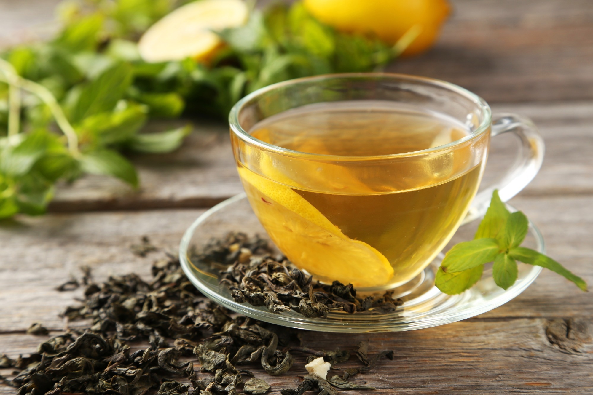
Plant polyphenols are micronutrients known to reduce inflammation and are abundant in green tea, which is both popular and widely available.
Green tea polyphenols are known as catechins, of which the most common is EGCG. While the role of EGCG in reducing inflammation has been studied, whether this could then improve iron status remains unknown.
About the study
Researchers from universities in Iowa and Indiana aimed to explore whether LPS-driven inflammation affects iron status and whether EGCG supplements can alleviate this impact.
They used lipopolysaccharide (LPS) to induce inflammation in albino laboratory rats and examine how inflammation and iron status are related. The experiment included 32 male rats assigned to one of four intervention groups (eight rats per group).
Before the treatments, all the rats were given iron-deficient food for two weeks so that they became iron deficient. Then, eight rats served as negative controls and remained on the iron-deficient diet.
Another eight were in the positive control group and were fed an iron-repletion diet. The first treatment group was given LPS, while the second group was exposed to LPS and EGCG as green tea powder.
Since LPS was administered to the treated rats through thrice-weekly intraperitoneal injections, the control rates were administered intra-peritoneal saline instead.
The research team measured the rats’ food intake and weight daily. After three weeks, the rats were anesthetized to collect blood, liver, and spleen samples.
Serum was extracted from blood samples to quantify iron status in terms of serum iron, hemoglobin, hematocrit, and ferritin, as well as inflammation markers – SAA, C-reactive protein (CRP), and interleukin 6 (IL-6). Total iron concentrations were derived from spleen and liver samples.
Findings
The rats weighed 61g on average when the study began and grew to 269g on average by the end, with no significant differences between the four groups.
Each rat consumed 10g of food per day across the four groups, while the EGCG-supplemented animals consumed about 5.8mg of green tea powder daily.
Negative control rats had lower hemoglobin concentrations than positive control and LPS treatment groups. However, positive control rats did not differ significantly from the two LPS groups. Similar results were seen for hematocrit levels, indicating that EGCG did not affect either of these iron status indicators.
While serum iron concentrations were not significantly lower in the negative control group, LPS-induced inflammation did reduce serum iron compared to the positive control group. Again, EGCG did not affect serum iron levels.
Liver iron levels were similar across all four groups. However, spleen iron levels were lowest in the negative control group; the three remaining groups showed similar concentrations.
A notable finding was the LPS increased the inflammatory marker SAA in the LPS group compared to the rats in the negative control group.
At the same time, ECGC supplementation lowered the SAA levels back to control levels. However, similar results were not seen for the other marker CRP. Surprisingly, IL-6 concentrations were lower in the LPS rats than in those who received EGCG supplementation.
Conclusions
This study is among the first to explore using dietary intervention to counteract inflammatory responses to improve iron levels and yielded mixed results. While LPS did reduce serum iron, EGCG did not counteract this effect.
However, EGCG supplementation did reduce the higher SAA levels caused by LPS, highlighting its anti-inflammatory effects.
The authors noted that they used lower LPS doses than previous studies on rats to reduce mortality. They also discussed the possibility of ‘iron trapping’ in which iron is trapped within the cells, reducing natural flows and thus iron concentrations in serum.
The inconclusive results for CRP could be due to its short half-life. Measuring CRP soon after LPS administration could lead to more significant findings.
Future studies can experiment with different LPS and EGCG dosages and study more biomarkers to shed light on the complex interactions between diet, inflammation, and iron status.