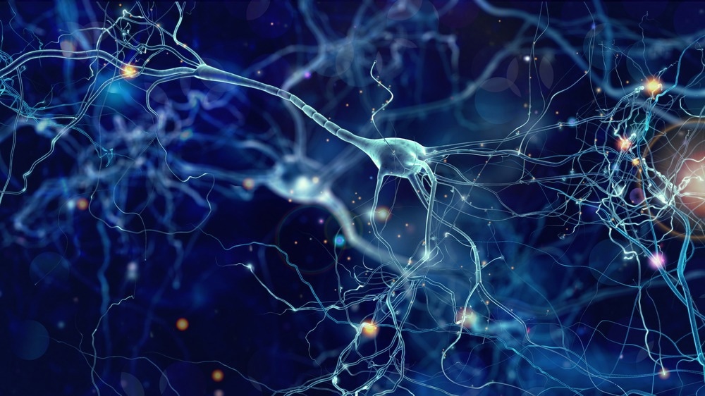
In a recent study published in Cell Stem Cell, researchers produced three-dimensional (3D) bioprinted human brain tissues, allowing for the creation of functioning neural networks that could simulate network activity in normal and pathological situations.
 Study: 3D bioprinting of human neural tissues with functional connectivity. Image Credit: whitehoune/Shutterstock.com
Background
Understanding neural networks in the human brain is critical for understanding brain health and disease. However, animal-based models cannot effectively reproduce the human brain's high-order data processing due to variations in cell composition, neural networks, and synaptic integration. 3D bioprinting provides a more accurate method for creating human brain tissues by physically repositioning hydrogels and live cells inside a physiologically complicated cytoarchitecture. However, bioprinting soft tissues, such as the brain, is of concern since soft biomaterials cannot sustain intricate 3D architectures or rigid gels.
About the study
In the present study, researchers developed a 3D bioprinting platform to manufacture tissues with defined human brain cell types in any desired dimension.
The team aimed to build layered neural tissues, including neural progenitor cells (NPCs) that generate connections inside and between the brain layers, maintaining the structure intact. They created a bioink for printing. They used fibrin gel to print the tissues. Methods for bioprinting include extrusion-based, laser-based, and droplet-based techniques. The extrusion three-dimensional bioprinting technique deposited gel in layers to simulate brain structures such as human cortex laminations.
The researchers selected a 50 mm thickness for every layer and built multi-layered tissues by placing the layers in a horizontal arrangement adjacent to one another. They designed 3D-printed brain tissues to be relatively thin but functional and multi-layered, with established cell compositions and desirable dimensions, and easily maintained and tested in a standard laboratory setting.
The researchers determined that 2.50 mg per mL fibrinogen and 0.50 to 1.0 U of thrombin were optimum concentrations for hydrogel formation, resulting in a gelation duration of 145 seconds, which allowed for 24-well plate printing. After six hours, most (85%) of the cells were viable and survived for seven days. The team created medial ganglionic eminence (MGE)-derived gamma-aminobutyric acid (GABA) and cortical (glutamate) progenitors from green fluorescent protein-expressing (GFP+) and GFP- human pluripotent stem cells (hPSCs) to investigate whether GABAergic interneurons and glutamatergic neurons form synaptic connections when inserted into printed tissues. Before printing, they combined the two progenitor populations in a 1:4 ratio to match the ratio of interneurons to cortical projection neurons in the cerebral cortex.
The researchers recorded electrophysiological data from tissues printed with GFP+ glutamatergic cortical progenitors, noncolored MGE GABAergic progenitors, and hPSC-derived astrocyte progenitors incorporated into glutamate neurons and GABA interneurons. The printed tissue was immunostained with an axonal marker, SMI312. They studied Alexander disease (AxD), a neurodegenerative disease caused by GFAP gene abnormalities, to investigate pathogenic mechanisms. They used live imaging of glutamate uptake by glutamate-sensitive fluorescent reporters (iGluSnFR) to investigate neuron-astrocyte interactions and neuron-glial connections in AxD.
Results
The printed neuronal progenitors developed into neurons within weeks, forming functional neural networks inside and across tissue layers. Printed astrocyte progenitors matured into astrocytes with complex processes to function in neuron-astrocyte networks. Conventional culture techniques could retain the 3D brain tissues, making them easier to investigate in physiological and pathological settings. Cell viability declined with rising concentrations of thrombin at 2.50 mg/mL fibrinogen concentrations but remained unaltered at a constant concentration of 0.50 U fibrinogen, and cells aggregated at increased fibrinogen levels.
The bioprinted neural cells matured and retained tissue form, with GFP-expressing cells in one band transforming into microtubule-associated protein 2 (MAP2+) neurons a week after printing. The printed tissue maintained a stable configuration where neural progenitors multiplied and built neural networks. The neuronal subtypes established functional networks within the bioprinted tissues, with hPSC-derived MGE cells expressing NK2 homeobox 1 (NKX2.1) and GABA and cortical progenitors positive for forkhead-box G1 (FOXG1) and paired box 6 (PAX6). The bioprinted neural tissue constructions promote the growth of cortical glutamatergic neurons and GABAergic interneurons.
The researchers utilized a high-concentration potassium chloride solution to print tissues containing neurons and astrocytes, demonstrating functional connections. The astrocytes expressed glutamate transporter 1 (GLT-1), indicating maturation. The printed cortical and striatal neuronal bands remained intact 15 days after printing, and GFP and mCherry neurites developed towards each other. The printed human brain tissues might replicate diseased processes, with AxD astrocytes exhibiting intracellular GFAP aggregation. By 30 days, MAP2+ neurons and GFAP+ astrocytes exhibited complex morphology and synapsin expression.
Conclusion
Overall, the study findings demonstrated the ability of 3D printing to generate functioning brain tissues for simulating network activity in normal and pathological settings. The bioink-created tissues establish functional synaptic connections between neuronal subtypes and neuron-astrocyte networks in two to five weeks. The 3D platform provides a defined environment for studying human brain networks in healthy and pathological settings; however, it has limitations, such as the softness of the gel and the 50mm thickness of the printed tissues.