In a recent study published in the United States (U.S.) Centers for Disease Control and Prevention (CDC)’s Emerging Infectious Disease Journal, a team of scientists reported the spread of the highly pathogenic hemagglutinin 5 neuraminidase 1 (H5N1) avian influenza clade 2.3.4.4b among cats and dairy cattle in the states of Texas and Kansas.
The highly pathogenic H5N1 avian influenza viruses have long been a threat to poultry and wild bird populations worldwide. They have also been a cause for concern in recent years because of their ability to infect various mammalian species. North America experienced an outbreak of the Eurasian strain clade 2.3.4.4b of the H5N1 virus that has continued into this year, with multiple spillover events reported among marine and terrestrial mammals.
The current avian influenza outbreak also poses more serious concerns since the clade has also been detected in severe cases of infections among humans in Chile and Ecuador. Recent reports from northern Texas, northeastern New Mexico, and southwestern Kansas reported a disease among dairy cattle that affected the quality and appearance of milk for a duration of approximately two weeks. Incidents of death were also reported from these states among domestic cats and wild birds, indicating a potential spillover of the avian influenza virus into cats and cattle.
About the study
In the present study, the researchers described the various characterization analyses performed to detect the highly pathogenic H5N1 avian influenza virus infecting domestic cats and cattle in the states of Texas and Kansas. These tests were performed at the Veterinary Diagnostic Laboratory at Iowa State University.
Milk and tissue samples were provided to the laboratory from cattle exhibiting clinical signs such as an abrupt decrease in milk production, thickening and yellowing of milk, and decreased rumination and food intake. The tissue samples were from cows that were either euthanized or died naturally. Additionally, the laboratory also performed postmortem analyses on two domesticated cats that had died in a dairy farm in north Texas after being fed milk from the sick cows.
Histopathology analyses were performed on the cerebellum, cerebrum, eye, heart, kidney, liver, lung, lymph node, and spleen obtained during the postmortem analysis of the cats. Additionally, paraffin-embedded tissues from the cats and cattle were processed for immunohistochemistry analyses using antibodies against the primary influenza A virus.
Milk samples diluted with phosphate-buffered saline, homogenates of brains, mammary glands, lymph nodes, spleen, and lungs, as well as samples of serum, rumen content, and ocular fluid, were processed for viral ribonucleic acid (RNA) isolation. Real-time reverse-transcriptase polymerase chain reaction (rRT-PCR) was performed to screen these samples for influenza A virus RNA, and only those samples that had cycle threshold values above 40 were considered positive.
Additional rRT-PCR methods were then used to analyze the H5 subtype and test for the H5 clade 2.3.4.4b in the samples that were positive for influenza A virus. Furthermore, the researchers also conducted genomic sequencing for two tissue samples from the cats and two milk samples. The data obtained was analyzed to assemble eight segments of influenza A virus sequences. Of these, the hemagglutinin and neuraminidase sequences were used for phylogenetic analyses.

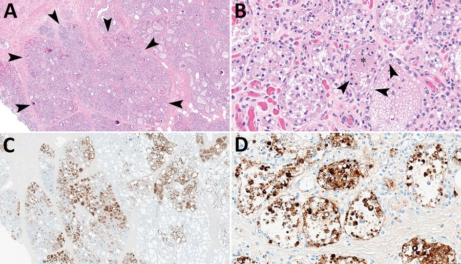
Mammary gland lesions in cattle in study of highly pathogenic avian influenza A(H5N1) clade 2.3.4.4b virus infection in domestic dairy cattle and cats, United States, 2024. A, B) Mammary gland tissue sections stained with hematoxylin and eosin. A) Arrowheads indicate segmental loss within open secretory mammary alveoli. Original magnification ×40. B) Arrowheads indicate epithelial degeneration and necrosis lining alveoli with intraluminal sloughing. Asterisk indicates intraluminal neutrophilic inflammation. Original magnification ×400. C, D) Mammary gland tissue sections stained by using avian influenza A immunohistochemistry. C) Brown staining indicates lobular distribution of avian influenza A virus. Original magnification ×40. D) Brown staining indicates strong nuclear and intracytoplasmic immunoreactivity of intact and sloughed epithelial cells within mammary alveoli. Original magnification ×400.
Results
The results indicated systemic illness, viral shedding in milk, and reduced milk production observed in dairy cattle were due to infection from a strain of the H5N1 influenza A virus. This highly pathogenic avian influenza virus also resulted in the death of approximately half the cats that had consumed the milk from the sick cows. Furthermore, the histopathological analyses revealed lesions in the tissue samples from cats similar to those observed in tissue samples obtained from cats that presumably got infected after eating wild birds with avian influenza infection.
The only tissues from the infected cows that were positive in the immunohistochemistry analysis using antigens against the influenza A virus were mastitic mammary gland samples. However, the tissue samples from both cats revealed microscopic lesions indicative of systemic viral infection, such as lymphocytic meningoencephalitis involving neuronal necrosis and vasculitis, lymphoplasmacytic chorioretinitis with necrosis of ganglion cells, and many more clinical findings. Additionally, the brain, heart, lung, and retinal samples were positive for influenza A virus immunoreactivity.
The phylogenetic analyses using the hemagglutinin and neuraminidase sequences also revealed high similarity between the sequences from the milk samples and those from the cats. These sequences were also highly similar to published sequences for H5N1 viruses of clade 2.3.4.4b.
Conclusions
Overall, the results showed that domestic cats and dairy cattle were susceptible to the highly pathogenic H5N1 avian influenza virus. Furthermore, viral shedding through milk increased the potential for the virus to get transmitted to other mammalian species. The recurrence of avian influenza outbreaks and the broadening of the host range are of growing concern, and surveillance of the virus in domesticated animal populations is essential to prevent the transmission of the virus across species.
Journal reference:
- Burrough, E., Magstadt, D., Petersen, B., Timmermans, S., Gauger, P., Zhang, J., Siepker, C., Mainenti, M., Li, G., Thompson, A., Gorden, P., Plummer, P., & Main, R. (2024). Highly Pathogenic Avian Influenza A(H5N1) Clade 2.3.4.4b Virus Infection in Domestic Dairy Cattle and Cats, United States, 2024. Emerging Infectious Disease Journal, 30(7). DOI: 10.3201/eid3007.240508, https://wwwnc.cdc.gov/eid/article/30/7/24-0508_article