Sponsored Content by GrundiumNov 25 2019
The utilization of digital microscopes to perform whole-slide imaging (WSI) provides multiple advantages to pathology, bioscience, and healthcare research. However, the ubiquity and simplicity of optical microscopes has hindered the adoption of digital microscopy methods.

This article outlines how new technology for whole-slide imaging and digitization is eradicating these entry barriers and allowing more organizations to reap the rewards of digital pathology.
Electronic data storage and transfer has revolutionized the way many industries and organizations operate. Biopsy samples are unable to be emailed and, as a result, professionals and researchers in biosciences and healthcare still frequently rely on the transfer of intricate and often delicate physical samples between locations for them to be analyzed properly.
This is particularly true in pathology, where the scarcity of pathologists is a serious problem and research institutions and hospitals often have to transport samples and microscope slides to external pathologists for analysis.1
Using slide-digitizing technology, these challenges have been partially addressed. Digital microscopy may be utilized to scan slides and generate a 2D or 3D composite digital image, which can then be distributed easily to other places that they can be analyzed properly by the relevant experts.

This ‘digital pathology’ has helped bioscience and healthcare professionals to overcome the chronic global shortage of pathologists. The process of obtaining an expert pathologist’s analysis can be made vastly more efficient by sending digitized microscope slides instead of physical specimens.
Furthermore, digitized slides can be stored, archived and retrieved far more easily than physical microscope slides, making sample data more readily available to pathologists, health professionals, and patients.
The slide-scanning technology, which makes digital pathology possible, has received endorsement from the FDA for primary diagnosis in surgical pathology in 2017,2 and has been around for more than 20 years. The adoption of these methods and technologies has been slow for multiple reasons, despite having clear benefits.
Among these, financial concerns are key: traditional optical microscopes, slides, and all they entail are a proven technique.3 High-throughput digital microscopy systems can be prohibitively expensive, and administrators at hospitals and research facilities can be understandably reluctant to invest in new technologies without having observed a proven return on investment.
Furthermore, there is an associated financial risk that new technology often needs investment in training for the people who will use it. Optical microscopes have such a long history and breadth of application that they have become fixtures in any laboratory.
Researchers, doctors, scientists, and pathologists who have trained using optical microscopes and work with them daily may be reluctant to adopt digital microscopy methods, particularly if there is a learning process associated with the new technology.

Lowering Barriers to Digital Microscopy
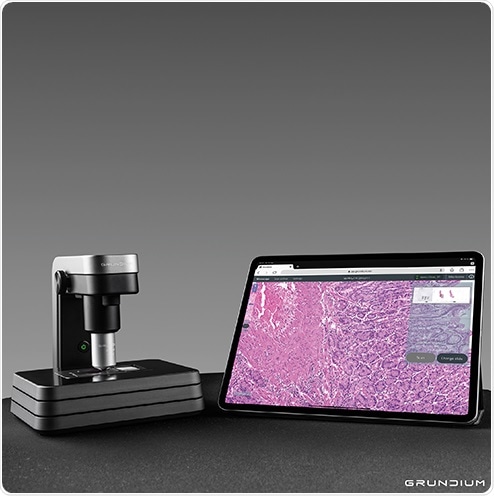
Using the Ocus, an extremely compact and portable WSI microscope, Grundium is tackling these barriers to adoption. What makes the Ocus unique is that its entire design has been considered carefully, specifically to lower barriers to the adoption of digital microscopy.4
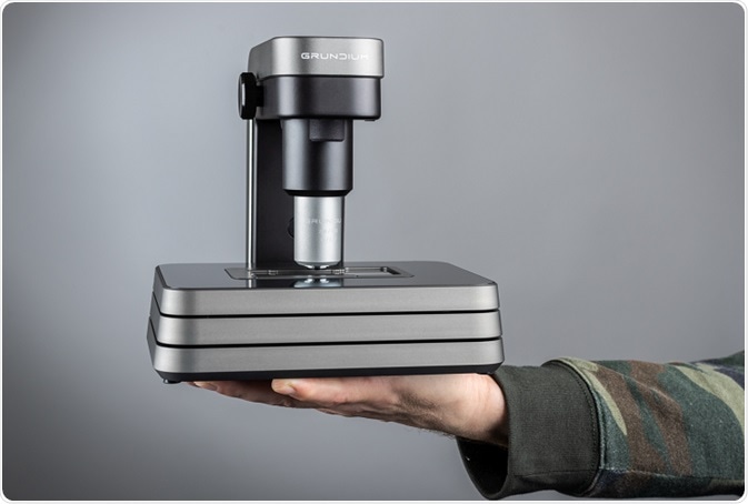
First, the Ocus is extremely small. Not just “benchtop” small, the 3.5 kg device is versatile and light enough to be carried from place to place, whether that means into the field to analyze botanical samples or the inside of a cryosection room in neuropathology.
With a water-proof carry case and battery pack, which fits easily into hand luggage, the Ocus can be employed to analyze samples anywhere at any time.5 The Ocus is also user-friendly to the point that it does not need training or practice to scan slides.
Retrieving scans is even easier. The Ocus includes 500 GB of internal storage but can export them automatically to a preferred storage location or cloud just as easily. The Ocus is easily accessible and can be remotely utilized via a web interface from any tablet, computer, or phone, with no specialist software to be installed or downloaded.
The choice to connect to the internet via onboard Wireless LAN or wired gigabit Ethernet means that it can be easily networked into any lab and utilized from anywhere around the globe.
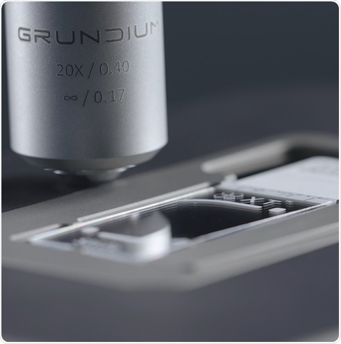
The Ocus is nothing less than a research-grade tool, despite its ease-of-use and size. Image sensors, high-performance optics, and LED illumination gather high-resolution images, which are quickly stitched together via a powerful onboard processor to supply 18 million-pixel composite images.

During the scanning process, automatic focus adjustment means that, when viewing the scan, the full scope of focal lengths may be looked at. Robust full-metal design and in-built vibration resistance mean that the Ocus is rugged enough to be employed anywhere and needs almost no maintenance.
The Ocus supports a “live view” in addition to scanning slides. This works similarly to a conventional microscope, but the Ocus may be remotely controlled, which allows pathologists to inspect samples remotely.
This is particularly helpful for assessing frozen sections. The Ocus can be kept in the frozen section lab where a technician prepares slides while its operator observes samples remotely from the comfort of his home office. The live view is the fastest method of viewing areas of interest and allows pathologists to look at slides in critical-use cases quickly.
In addition to allowing digital archiving of slides, enhancing turnaround times, and sharing digitized samples with specialists quickly, WSI could be vital in the future. Digitizing scans means that they can be analyzed by artificial intelligence systems to speed up diagnoses.
Grundium are hoping that the Ocus will play a key role in a paradigm shift away from traditional optical microscopes and time-consuming distribution of physical slides towards simpler, quicker, and more streamlined analysis via WSI.

References and Further Reading
- Petriceks, A. H. & Salmi, D. Trends in pathology graduate medical education programs and positions, 2001 to 2017. Acad. Pathol. 5, (2018).
- FDA allows marketing of first whole slide imaging system for digital pathology | FDA.
- Randell, R., Ruddle, R. A. & Treanor, D. Barriers and facilitators to the introduction of digital pathology for diagnostic work. doi:10.3233/978-1-61499-564-7-443
- Grundium Ocus.
- Grundium - Collaboration and Off-Lab Work.
About Grundium
The Grundium Ocus does away with physical slide transfers between the clinic and the lab and enables live telepathology consultations between surgeons and pathologists.
The Grundium Ocus is a monumental leap in digital pathology. It is a precision tool, and is small and affordable enough to be on every medical professional’s desk. It is truly portable and can be brought anywhere. Wireless connectivity means telepathology is now possible practically anywhere on the planet.
Key Benefits
The Grundium Ocus is a game changing enabler of personal digital microscopy. It brings the global pool of pathology experts to you and it makes looking at sample slides convenient even off-lab.
The Grundium Ocus is your personal microscope scanner to make daily work faster and easier.
Sponsored Content Policy: News-Medical.net publishes articles and related content that may be derived from sources where we have existing commercial relationships, provided such content adds value to the core editorial ethos of News-Medical.Net which is to educate and inform site visitors interested in medical research, science, medical devices and treatments.