Modern high-content screening microscopes provide fast, automated imaging of entire microtiter plates by capturing fixed positions within each well. This approach is particularly suited for in-vitro cell culture assays or other applications with evenly distributed phenotypes.
However, the limited field of view (FOV) of high-magnification objectives may not cover the entire region of interest, presenting challenges when studying large specimens or rare events.
As a result, researchers are frequently limited to lower magnification acquisition, resulting in low-resolution data or the omission of features of interest in many wells.
This article explains how Bruker's Acquifer Imaging Machine (IM), with its Plate-Viewer software, addresses these limitations and provides a semi-automated approach for advanced, supervised feedback microscopy experiments.
Automated experimental approaches
For tissue-specific imaging in large specimens (e.g., zebrafish), an approach is needed that can automatically identify and zoom in on the tissue or organ of interest.
Fully automated tissue detection and imaging often demand the development of sophisticated image processing routines. Therefore, each project needs careful balancing between software development time and project size.
Semi-automated approaches offer an ideal compromise, as they only require minimal user interaction and no custom algorithm development. Even complex or variable structures that would require extensive development of image detection routines can be readily handled via this simpler alternative. This approach is beneficial for screening projects as they start instantly, saving time and resources.
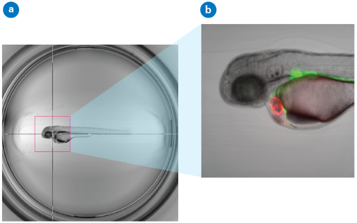
Figure 1. Illustration of Click-Tool functionality. (a) Zebrafish embryo imaged with a 2x objective and visualized in Plate-Viewer. The crosshair is centered on the heart region in a three-day-old embryo of the epi:GFP;myl7mR transgenic line. The red bounding box indicates the field of view of a 10x objective used for subsequent high-resolution imaging. (b) Single Z-plane of a high-resolution dataset is automatically acquired on Acquifer IM. Image Credit: Bruker Nano Surfaces and Metrology
Plate-Viewer software provides a semi-automated approach for supervised feedback microscopy. Low-magnification pre-screen data of a full microtiter plate is visualized according to the plate layout, offering a rapid and intuitive overview.
The integrated “click-tool” feature enables assay experts to choose regions of interest (ROIs) for each well.
Additionally, built-in “template matching” algorithms make it easy to automatically locate a wide range of target structures, from complex reporter expression patterns to specific morphological features or rare events in each well.
With Acquifer IM, high-resolution data is automatically captured from selected regions based on your predefined settings. The results are then displayed in Plate-Viewer, offering a clear and intuitive way to explore the data.
Plate-Viewer is packed with features designed to make scientists' workflows smoother and more efficient:
- Select ROIs for automatic imaging with just a click or by using template matching.
- Preview FOVs at higher magnifications using an adjustable bounding box.
- Visualize and browse even large-scale screening datasets with ease.
- Seamlessly navigate through complex multidimensional datasets.
- Enhance manual inspection by applying LUTs and overlaying channels.
- Adjust basic image parameters to bring out finer details.
- Integrate external image processing tools through a convenient plug-in interface.
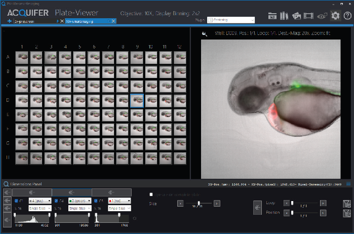
Figure 2. Overview of the Plate-Viewer functionalities. Visualization of identical planes of all available wells within a microtiter plate (left). Preview of the currently selected well coordinate (right). Control panels in the top right corner enable the generation of colored overlay images and the display of selected channels. Control panels in the lower left corner allow channel-specific adjustment of the histogram and the choice of navigation mode through multidimensional datasets (e.g., slice, loops, or subpositions). Image Credit: Bruker Nano Surfaces and Metrology
Intuitive solution for high-content screening
Acquifer IM offers a powerful solution for supervised feedback microscopy experiments, seamlessly integrating with Plate-Viewer software to simplify complex high-content screening studies.
Using a semi-automated approach, this advanced tool makes tissue-specific imaging of intricate or variable structures in large specimens easier and more reliable for scientists.
About Bruker Nano Surfaces and Metrology

Bruker’s suite of fluorescence microscopy systems provides a full range of solutions for life science researchers. Their multiphoton imaging systems provide the imaging depth, speed and resolution required for intravital imaging applications, and their confocal systems enable cell biologists to study function and structure using live-cell imaging at speeds and durations previously not possible. Bruker’s super-resolution microscopes are setting new standards with quantitative single molecule localization that allows for the direct investigation of the molecular positions and distribution of proteins within the cellular environment. And their Luxendo light-sheet microscopes, are revolutionizing long-term studies in developmental biology and investigation of dynamic processes in cell culture and small animal models.
Sponsored Content Policy: News-Medical.net publishes articles and related content that may be derived from sources where we have existing commercial relationships, provided such content adds value to the core editorial ethos of News-Medical.Net which is to educate and inform site visitors interested in medical research, science, medical devices and treatments.