Introduction
Neuroplasticity and Cortical Reorganization
Evidence for Improved Sensory Performance
Mechanisms and Constraints of Compensation
Emerging Research and Future Directions
References
Further reading
What happens in the brain when sight or hearing fades, and why do some remaining sensory abilities become sharper?
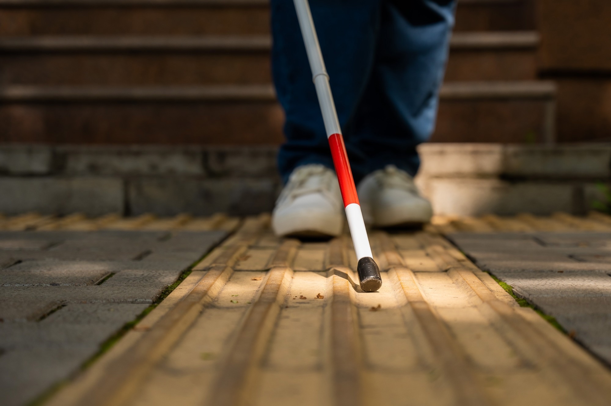 Image Credit: Reshetnikov_art / Shutterstock.com
Introduction
"Sensory compensation" describes neural and behavioral adaptations that support interaction with the environment after impairment of a primary sensory modality. Losing vision or hearing, for example, can improve performance on certain tasks that rely more on the remaining senses; however, these benefits vary by individual and task. Compensation is often incomplete, and sensory loss can continue to limit everyday functioning.1,2
Two related forms of plasticity are "cross-modal recruitment," in which sensory cortices receiving reduced input respond to information from other modalities, and "compensatory plasticity," which refines the functional circuits of the spared cortices.1,10 The extent and behavioral consequences of these changes vary with developmental timing, the severity of sensory loss, and experience, including sensory training and language use.2
Studying these adaptations can inform rehabilitation and sensory restoration, though researchers caution against assuming that all cortical changes produce functional benefits. Most human evidence discussed here concerns blindness and hearing loss.1,2
Neuroplasticity and Cortical Reorganization
Cortical development and the timing of sensory deprivation influence the pathways available for adaptation. During embryogenesis, thalamocortical (TC) axons, which carry information from the thalamus to the cortex, contribute to the development and differentiation of primary visual (V1), auditory (A1), and somatosensory (S1) cortical areas, alongside intrinsic developmental mechanisms.3
The consequences of sensory deprivation differ depending on whether sensory input is removed before or after TC axons invade the cortical plate, the developing layer that forms much of the cerebral cortex.3 In animal models with deprivation after TC axons begin entering the cortical plate, adaptations can involve altered intracortical communication and the unmasking or strengthening of existing connections.3
For instance, neonatal bilateral enucleation (removal of both eyes) in rats has been associated with a 20% expansion of cortical whisker representation. Separate observations describe recruitment of the rostral visual cortex for whisker-driven inputs.3
In models with deprivation before TC axons enter the cortical plate, including ferrets and hamsters, experimental manipulations can alter subcortical pathways. In ferrets, lesions involving the auditory midbrain and other sensory targets redirected retinal axons to the auditory thalamus, producing visual responses and V1-like orientation maps in A1. These combined lesion experiments should be distinguished from naturally occurring sensory loss, and their outcomes vary with the species and manipulation.3,7
In humans, neuroimaging studies show that blind individuals recruit occipital cortical areas during nonvisual tasks. Early-onset blindness is associated with occipital activity during haptic tasks, such as Braille reading, and auditory tasks, such as sound localization.1,2
Sensitive developmental periods influence these cross-modal recruitment patterns, and their timing varies across circuits and functions. Plasticity also occurs in adulthood; therefore, no single childhood age range defines its limits.1–3
 Image Credit: Kateryna Kon / Shutterstock.com
Some individuals with profound sensory loss show "supra-normal" performance on specific tasks involving the remaining senses. In early-blind cohorts, for example, this includes finer tactile spatial resolution and superior sound localization.2
Under monaural conditions (one ear plugged), a subgroup of early-blind individuals has demonstrated better sound localization than sighted controls. Such findings come from controlled experimental conditions and do not establish a general advantage across everyday listening situations.2
Superior verbal memory has also been reported in blind individuals, while occipital activation has been associated with performance. This cognitive advantage should be distinguished from increased sensitivity of a remaining sense.2
Some deaf individuals perform better than hearing controls on tasks involving peripheral visual motion and allocate more attention to the visual periphery. Early experience with sign language can also affect visual processing; therefore, researchers must distinguish language experience from auditory deprivation.2
These compensatory benefits are task-specific. Better peripheral performance does not establish uniformly better vision, and comparisons must account for the visual task, attentional demands, as well as participants' sensory and language histories.2
How Blind People See With Sound… with @MollyBurkeOfficial!
Mechanisms and Constraints of Compensation
At the cellular level, cross-modal compensation can involve synaptic modifications and long-range modulatory signaling. Using the Caenorhabditis elegans (C. elegans) model, loss of mechanosensory input reduces activity in touch neurons and decreases secretion of the neuropeptide FLP-20, disinhibiting inhibitory glutamatergic transmission in the olfactory circuit to improve olfactory acuity.5
In mammals, neuromodulators can also influence communication between sensory circuits. In rats, visual deprivation increases serotonergic signaling in the somatosensory cortex, strengthening layer 4-to-layer 2/3 excitatory glutamatergic connections.5
In neonatal mice, visual or somatosensory deprivation can reduce oxytocin secretion from the hypothalamus, weakening glutamatergic transmission to superficial layers of the other sensory cortex and reducing circuit output. This example shows that deprivation can also weaken processing in a spared sensory system.5
Metaplasticity, a change in the conditions required for subsequent synaptic strengthening or weakening, offers another explanation for adaptation. The Bienenstock-Cooper-Monroe (BCM) model proposes that reduced activity lowers the synaptic modification threshold (θM), making long-term potentiation easier to induce.
Evidence reviewed for V1 supports input-specific strengthening of intracortical synapses that requires N-methyl-D-aspartate (NMDA) receptor activity. This mechanism differs from synaptic scaling, which can occur without NMDA receptor activation.1
In rodent experiments, as little as two days of visual deprivation can strengthen lateral intracortical connections in V1 superficial layers, whereas A1 feedforward connections from the ventral medial geniculate body to layer 4 show strengthening after seven days of dark exposure.1 Importantly, these observations concern synaptic changes in experimental animals and do not define a universal timetable for human sensory improvement.1
The interpretation of cortical remapping remains unclear. Many apparent changes reflect stronger use of pre-existing circuitry with relevant computational capacities, with conflicting opinions on whether cortical regions acquire entirely new functions after sensory loss.6
In hearing loss, researchers have observed strengthening and weakening of existing connections, including top-down pathways carrying influences from higher-order brain regions. Cross-modal changes in partial and unilateral hearing loss have also been reported.6,7
 Image Credit: KinoMasterskaya / Shutterstock.com
Emerging Research and Future Directions
Longitudinal studies are needed to establish when cross-modal changes develop, how they relate to behavior, and whether they change after treatment. During these studies, brain stimulation can help test whether recruited cortical regions contribute to performance.
For example, transcranial magnetic stimulation applied to the occipital cortex can disrupt Braille reading in blind readers, supporting this region's functional contribution to the task. These experiments establish causal involvement in task performance, though therapeutic applications require separate evidence.2
Sensory substitution offers another way to investigate how the brain uses information from remaining senses. Haptic sensory substitution devices (SSDs) provide information usually obtained through another sense. The BrainPort device translates digital video into electrotactile patterns on the tongue, with reported experimental task performance corresponding to visual acuity of approximately 20/800.8 This measures performance using substituted information, rather than restoration of ordinary eyesight, with performance varying by task, training, and device configuration.8
Studies of partial hearing loss illustrate why neural and behavioral outcomes must be assessed separately. In mild-to-moderate age-related hearing loss, high-density EEG has shown significantly earlier P1, N1, and P2 latencies over a right temporal scalp region during visual tasks.9
A separate 2026 study examined 25 adults aged 18-78 with varying high-frequency hearing sensitivity and normal low- and mid-frequency hearing. Poorer high-frequency hearing was associated with larger visual N1 amplitudes and longer P2 latencies. Source analysis, which estimates the brain regions contributing to scalp-recorded EEG signals, linked the larger N1 response to the right auditory cortex.10
Treatment studies examine whether these neural responses change when auditory input improves. In the hearing-aid study, researchers reassessed 21 participants after 6 months of bilateral hearing aid use. Right temporal VEP latencies shifted toward values observed in the normal-hearing comparison group, and source estimates suggested reduced visual recruitment of auditory regions. These findings support treatment-associated changes in adult cross-modal activity, but the study was not randomized and lacked a comparison group retested at six months. It cannot establish complete reversal in every individual or attribute all changes exclusively to hearing aid use.9
Together, these findings support selective adaptation after sensory loss. Measured performance remains necessary to establish functional compensation, while the contributions of residual sensory function, developmental history, and training vary across populations and tasks.1,2,4,10
References
- Ewall, G., Parkins, S., Lin, A., et al. (2021). Cortical and Subcortical Circuits for Cross-Modal Plasticity Induced by Loss of Vision. Frontiers in Neural Circuits 15. DOI: 10.3389/fncir.2021.665009. https://www.frontiersin.org/journals/neural-circuits/articles/10.3389/fncir.2021.665009/full
- Merabet, L. B., & Pascual-Leone, A. (2010). Neural reorganization following sensory loss: the opportunity of change. Nature Reviews Neuroscience 11(1); 44–52. DOI: 10.1038/nrn2758. https://www.nature.com/articles/nrn2758
- Mezzera, C., & López-Bendito, G. (2016). Cross-modal plasticity in sensory-deprived animal models: From the thalamocortical development point of view. Journal of Chemical Neuroanatomy, 75; 32-40. DOI: 10.1016/j.jchemneu.2015.09.005. https://www.sciencedirect.com/science/article/pii/S0891061815000721?via%3Dihub
- Aguiar, P. V., Preman, J., & Paul, B. T. (2025). Cross-modal neuroplasticity in partial hearing loss: a mini-review. Frontiers in Neuroscience 19. DOI: 10.3389/fnins.2025.1627888. https://www.frontiersin.org/journals/neuroscience/articles/10.3389/fnins.2025.1627888/full
- Rabinowitch, I. & Bai, J. (2016). The foundations of cross-modal plasticity. Communicative & Integrative Biology 9(2); e1158378. DOI: 10.1080/19420889.2016.1158378. https://www.tandfonline.com/doi/full/10.1080/19420889.2016.1158378?scroll=top&needAccess=true
- Makin, T. R., & Krakauer, J. W. (2023). Against cortical reorganization. eLife 12. DOI: 10.7554/elife.84716. https://elifesciences.org/articles/84716
- Kral, A., & Sharma, A. (2023). Crossmodal plasticity in hearing loss. Trends in Neurosciences 46(5); 377-393. DOI: 10.1016/j.tins.2023.02.004. https://www.cell.com/trends/neurosciences/fulltext/S0166-2236(23)00045-0?_returnURL=https%3A%2F%2Flinkinghub.elsevier.com%2Fretrieve%2Fpii%2FS0166223623000450%3Fshowall%3Dtrue
- Eagleman, D. M., & Perrotta, M. V. (2023). The future of sensory substitution, addition, and expansion via haptic devices. Frontiers in Human Neuroscience 16. DOI: 10.3389/fnhum.2022.1055546. https://www.frontiersin.org/journals/human-neuroscience/articles/10.3389/fnhum.2022.1055546/full
- Glick, H. A., & Sharma, A. (2020). Cortical Neuroplasticity and Cognitive Function in Early-Stage, Mild-Moderate Hearing Loss: Evidence of Neurocognitive Benefit From Hearing Aid Use. Frontiers in Neuroscience 14. DOI: 10.3389/fnins.2020.00093. https://www.frontiersin.org/journals/neuroscience/articles/10.3389/fnins.2020.00093/full
- Paul, B. T., Srikanthanathan, A., Daien, M., & Dimitrijevic, A. (2026). Association between high-frequency hearing sensitivity and visual cross-modal plasticity during active visual stimulus processing. Journal of Neurophysiology 135(1); 249-260. DOI: 10.1152/jn.00214.2025. https://journals.physiology.org/doi/full/10.1152/jn.00214.2025
Further Reading
Last Updated: Nov 17, 2026
 Does coffee truly boost memory and focus or is it all hype?