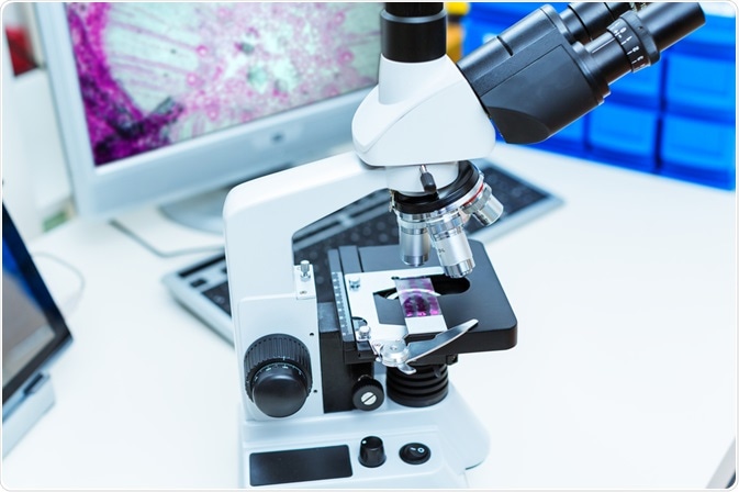
Digital pathology is being embraced by a growing number of laboratories around the world. The practice involves scanning whole slide images of tissue samples, analyzing them, uploading them into a database, and sharing them with labs globally.
 Image Credits: science photo / Shutterstock.com
The Food and Drug Administration has approved digital pathology for use as a tool of primary diagnosis in the USA. However, while digital pathology is being increasingly adopted across the nation, only a small portion of laboratories have fully implemented the system for primary diagnosis.
The idea of digital pathology has been around for decades, although it has only become popular recently as advances in technology have allowed it to begin to reach its full potential.
Digital pathology relies on computers with high processing powers, an internet connection, and sophisticated technology, such as artificial intelligence (AI), to give it its full range of analytical, automation, library, and communication capabilities. Because of this, the adoption of digital pathology is still gaining traction, as the technologies that support it are still growing.
Scientists need proof of the benefits of digital pathology in order to justify the expense of setting it up. While costs have come down in recent years, it is still an investment that needs hard evidence to back up its claims.
One of the major benefits of digital pathology is that it maximizes efficiency. It does this in a number of ways, below we explain how.
How digital pathology maximizes efficiency
Firstly, digital pathology uses AI to automatically select regions of interest in the scanning tissue slides. Usually, it is the responsibility of the pathologist to use their knowledge and training to manually select these regions. Digital pathology can carry out this task much quicker and to a higher degree of accuracy. It also frees pathologists to work on different tasks, leaving the menial ones to technology.
In addition, digital pathology can employ AI to automatically analyze the tissue samples. This can speed up diagnosis timeframes, and again, make pathologists available to spend their time on other tasks. Digital pathology can also be used to check the diagnoses made by pathologists if the lab still chooses to rely on human workers for this task. Overall, this can boost efficiency by preventing errors, eliminating the time and resources spent correcting for these errors.
A unique way digital pathology helps maximize efficiency is that it brings together experts from all over the world to work collaboratively without the expense of time or money on traveling to work together in person.
Furthermore, coworkers do not need to be in the lab at the time a tissue sample was investigated to give their input, they can look at it at a time that suits them, reducing the time spent waiting for colleagues to assess each other’s slides.
The ability to bring together pathologists in this way is having a huge impact on how quickly scientific breakthroughs can be made. By facilitating this collaborative work style, scientists can gain access to richer data sets as well as different analytical viewpoints, helping them to expand their knowledge on the pathology of a disease, leading to the faster development of new drug therapies and diagnostic methods.
The problem is that as digital pathology is still relatively new, there is currently a limited number of published studies that support the true benefit that digital pathology delivers. This is acting as a major barrier to adoption, as scientists have limited data on how digital pathology can realistically enhance efficiency in their lab.
Fortunately, studies are beginning to be published that demonstrate the efficiency of the method over conventional techniques.
Studies investigating the efficiency maximizing benefit of digital pathology
Last year, a team of researchers published a study that shows that digital pathology potential to significantly impact the field of pathology. In comparison to imaging glass slides using conventional methods, whole slide imaging utilized by digital pathology has been proven to be more efficient.
In a study published last year in the journal Modern Pathology, scientists revealed that, in comparison to conventional microscopy, complete digital pathology utilizing whole slide images had great levels of efficiency.
The study found that the two tests had high levels of diagnostic equivalency and that digital pathology operation resulted in increases in efficiency and operational utility. More studies like this are required to not only test the use of digital pathology so it can continue to advance and improve but so that pathologists can be assured of the benefit of the system to encourage its continued widespread adoption.
Overall, digital pathology increases efficiency in pathology labs in a number of ways. It automates the selection of the region of interest, as well as conducting analyses with AI. This enables it to speed up processes, and free up pathologists to work on other tasks, boosting efficiency. It also reduces time and resources lost to correct mistakes.
Finally, and most importantly, is that it allows scientists to work collaboratively regardless of their location, helping to speed up drug development and diagnosis. To enable digital pathology to continue to grow, more published data is needed to illustrate its benefit.
Sources:
Hanna, M., Reuter, V., Hameed, M., Tan, L., Chiang, S., Sigel, C., Hollmann, T., Giri, D., Samboy, J., Moradel, C., Rosado, A., Otilano, J., England, C., Corsale, L., Stamelos, E., Yagi, Y., Schüffler, P., Fuchs, T., Klimstra, D. and Sirintrapun, S., 2019. Whole slide imaging equivalency and efficiency study: experience at a large academic center. Modern Pathology, 32(7), pp.916-928. https://www.nature.com/articles/s41379-019-0205-0
IntelliSite Pathology Solution. Available at: www.fda.gov/.../intellisite-pathology-solution-pips-philips-medical-systems
Pantanowitz, L., 2010. Digital images and the future of digital pathology. Journal of Pathology Informatics, 1(1), p.15. https://www.archivesofpathology.org/doi/full/10.5858/arpa.2013-0093-CP
Further Reading
Last Updated: Jun 17, 2020