An interview with Patrick Myles from Huron Digital Pathology, discussing the digitization of whole mount slides and its impact on the field of neurobiological research.
How important are advances in imaging and IT to the progress of neuroscience research?
The ability to image the brain, so that it can be visualized and shared, is crucial to revolutionizing neuroscience research. Even a few years ago, what we're able to do today wasn't possible. The combination of being able to digitize whole mount slides and the availability of low cost storage and computer horsepower that has helped to make that a reality.
What role does whole mount digital slide imaging play in this field and how does this tool compare to previous methods?
In the simplest form, it gives a bigger, more complete view of the brain tissue. It also allows for better visualization of that imagery. As well as being able to look at an entire brain section, through 3-D reconstruction, you can model the brain and share that image data with other researchers. That will spawn new areas of research, new insights and new discoveries.
As for how it compares to the previous methods, ten years ago everything was done with a microscope. Even five years ago, the digitization of slides was available, but smaller sections of a brain would be taken and then scanned on standard sized slides. They would then reconstruct those to recreate the whole mount, which was incredibly time-consuming and problematic.
The ability to now scan an entire brain section at high magnification and do so in a decent amount of time is what is suddenly leading to all of these amazing possibilities for accelerating the research.

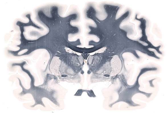
Whole Mount Brain - 5" x 7" at 20X. Image credit: Huron Digital Pathology.
Please can you give a brief overview of the TissueScope LE scanner? How many brain sections can be automatically scanned using this tool?
Our platform is the TissueScope LE and it's a brightfield scanner. It can scan any size of slide from 1 by 3 inches, all the way up to 6 by 8 inches and up to 40x magnification. The way that our slide holders are set up with our standard TissueScope LE, is such that you can scan twelve 1 by 3 inch slides or a single whole mount slide.
Typically, a brain section might by 5 by 7 inches, for example. That's our standard scanner, but we also have its older brother, the LE 120, which is able to load 10 slide holders so that you're scanning 10 brain sections automatically, without intervention.
For example, you can set it up overnight and come back in the morning to find the scans completed. What this effectively allows the researchers to do, is to scan up to potentially thousands of whole mount sections per brain.
How high is the resolution and what other benefits does the tool provide?
The magnification is 40x. The smallest feature size is 0.25 microns. From my experience and the experience of our customers, that is plenty of resolution. Typically, even 10x is more than enough and 20x is certainly more than adequate.
What we bring to the table is a lot of flexibility. For example, there may be some researchers in neuroscience that are interested in scanning mouse brains, primate brains or perhaps post-mortem human brains. With our system, they can go from one type of research to the next without being hindered by the size of the slide. That's the biggest benefit…the flexibility.
There are a lot of companies out there that scan and share slides, but we provide a solution that allows the flexibility to image where it otherwise wouldn't be possible. For neurologists who want to do whole mount, we're a good company to work with because we've been doing this for a long time.
Huron Digital Pathology Innovative Slide Holder
Who has access to the TissueScope LE scanner?
What's exciting is that we've been providing this type of scanner for about five years, although more from a custom development standpoint.
One of the benefits of our LE platform is that it's a standard product and it's also accessible in terms of pricing, meaning a much broader range of researchers can now afford to scan whole mount slides and complete their research in a reasonable amount of time.
It accelerates their research and it's not just limited to one or two researchers in the world. Now, potentially hundreds of researchers around the world can now carry out this type of work. It's pretty exciting.
How data intensive is this tool and how are advances in low cost storage and computing horsepower impacting upon what is possible?
As well as our innovation in terms of the imaging scanning side, we're fortunate that the price of storage and the price of computer horsepower is continually coming down. Now, we're at a stage where it's quite viable to be able to scan literally thousands of sections and reconstruct and share that image data.
This scanning modality does push the limits of imaging horsepower. Every 6 months we see improvements in the costs of storage or the speed at which we can transfer data. It's all coming together now as an increasingly viable solution. It's already a viable solution today, but to be able to scan at 10x, perhaps 20x at whole mount is more than manageable. Over time, 40X will be the norm.
What it comes down to is how much storage capacity is required. It was only 10 years ago that somebody would talk about a terabyte of storage as being a huge amount of data, whereas now we're talking hundreds of terabytes. Soon a petabyte of data will be a normal, everyday occurrence.
These things go hand-in-hand and we work very hard to take advantage of those increases. The other aspect is the way that we save our files (what they call TIF files, which is a non-proprietary file format) in what is called a pyramidal file format. It's analogous to what's used with Google maps. From an image serving standpoint or a user standpoint, it's quite efficient to be able to access these images from just about anywhere in the world.
What impact do you think the broad adoption of whole mount brain imaging and related image analysis will have on research into neurological diseases?
First off, I have a personal vested interest in wanting to see some of these advances. My mother has Parkinson's disease and my mother-in-law has some early stages of dementia. I'm very keen on being able to provide these kind of tools to researchers so that they can accelerate the research that is needed for these and other diseases. In general, the ability to image the brain, to visualize it and then fuse those images together with other modalities such as X-ray or MRI, I think will help to accelerate the research.
The beauty of this is that the more researchers that have access to this imagery, the more insight there will be, which can only accelerate the research and the discoveries made. We already see it with some of our customers who, after having access to our scanner for even just a couple of months, have gained insights they just did not have when they were looking at an image through a microscope.
They'll be able to make connections and then share those images with their colleagues, who will see the same thing but may have other insights. There's a crowd sourcing aspect to it. It's not unlike what happens in a social media context with the sharing of images, videos or insights, for example. The more that we understand it and the more eyes that are on it, the more likely it will be that we'll suddenly make amazing discoveries that weren't possible before.
What are Huron Digital Pathology's plans for the future?
Our immediate plan and focus is to help our customers adopt whole mount. Whole mount presents a number of challenges. Preparing the tissue represents one challenge, for example, because these aren't standard size tissues. Our goal is to make whole mount scanning a lot easier to do.
Even though we're a scanner and image management company and not a tissue processing company, we work hard to help our customers be able to find a solution and do what they need to do. We happen to have good connections with various researchers around the world that are doing the same, so when another researcher comes along, we can make those connections.
Over time, what we'll do is start to provide tools that will allow for much easier sharing of the images created. It's one thing to be able to create these wonderful, highly detailed images, but it's another to be able to share them and we're starting to offer solutions for that.
We also have a strong capability in the fluorescence side of the imaging spectrum. We want to be able to eventually add some of those modalities to our very easy-to-use LE platform. What you would then have are tools that can do both brightfield and fluorescence, so you open up even more avenues for discovery.
Where can readers find more information?
About Patrick Myles
 Patrick became President of Huron Digital Pathology in June 2015. He joined the company in 2014 as Executive Vice President, Business Development.
He brings to the company 18 years of experience at Teledyne DALSA, an international manufacturer of digital imaging components with $250 million in annual revenue.
There, he was responsible for corporate business development, seeking out and evaluating opportunities to drive organic and inorganic growth.
Prior to DALSA's acquisition by Teledyne in 2011, Patrick had responsibility for investor relations, marketing, corporate communications, and served as corporate secretary.
Patrick has a Bachelor of Arts degree from the University of Waterloo and an MBA from Wilfrid Laurier University. He has served as a board member of the United Way Kitchener Waterloo and Area, the University of Waterloo Magazine advisory committee, and the Ontario Chapter of the Canadian Investor Relations Institute.