Sponsored Content by AlveoleFeb 18 2019
News-Medical speaks to Marie-Charlotte Manus from Alvéole, to find out about a new technology called PRIMO that improves the physiological relevance of cells in culture by giving researchers the ability to customize the cellular microenvironment.
What is the cellular microenvironment and how does it influence cellular mechanisms?
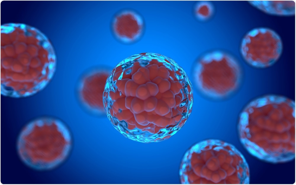
The cellular microenvironment is defined as the environment surrounding cells. Like organisms, cells are sensitive to their environment and respond to it. Within an organism (in vivo), this microenvironment is highly complex and dynamic, containing other cells with which one cell will interact, various molecules, and a specific stiffness (think of bones compared to the skin for instance), amongst others.
 m63085 | Shutterstock
These mechanical and biochemical factors are sensed by the surrounding cells, inducing a response. In addition, the microenvironment can impact cell proliferation, differentiation, and development.
What is micropatterning and how is this technique used by cell biologists?
Micropatterning is an in vitro technique which involves creating protein patterns on a substrate, upon which living cells can adhere. This allows researchers to mimic some in vivo conditions or to study the intracellular mechanism in a very controlled and reproducible way.
To give you some more precise examples, researchers can force cells to be confined on a specific shape which could mimic the shape it would have adopted in an organism. This technique can also be used to create lines of molecules on which cells can move and migrate allowing researchers to study and better understand the mechanism involved in this process.
Micropatterning can also allow researchers to isolate and study single cells, but also to place one cell to a specific distance from another cell and see if they interact and how.
Why are current micropatterning techniques considered to be laborious and over complicated?
Actually, it is the optimization of the right protein micropatterns which is a laborious process.
Using current techniques, you would need to create a sort of stencil (called a mask) with specific shapes on it and use this to create your protein micropattern. But, if for some unknown reason, the shape you used doesn’t suit the cells you are studying, you need to create a new protein micropattern and therefore to create a new stencil or order already micropatterned substrates.
It is for this reason that we developed PRIMO, a technology that allows you to draw the shape you want to use and simply project it onto the substrate. The system allows you to create a micropattern within minutes. If you need to use another pattern, you simply draw a new shape and project it.
 Spectral Designs | Shutterstock
Please can you describe the PRIMO micropatterning platform in more detail?
The PRIMO technique is based on LIMAP technology (Light Induced Molecular Adsorption of Proteins) and combines a maskless and contactless photolithography system (PRIMO) controlled by a dedicated piece of software (named “Leonardo”) and a specific photoactivatable reagent (PLPP).
The combined action of UV and PLPP makes it possible to generate, in only a few seconds, any multi-protein pattern on standard cell culture substrates.
A few months ago, we updated our technology so that it now goes further, not only controlling the biochemistry of the microenvironment but also enabling researchers to control the topography and stiffness of the microenvironment which can have a huge impact on cell behavior and function.
As a photopatterning system, PRIMO can be used for microfabrication to create microstructured substrates and recreate the shape of in vivo microenvironments in 3D. The UV projected on a photosensitive material will react with it (polymerization) and stiffened it accordingly to the projected shape.
Why is the physiological relevance of cells in culture important, and how does PRIMO provide this?
Every cell within a living organism is constantly interacting with its environment. This means that if you study them in an environment that is completely different to the one in which they would naturally evolve in “real life” in an organism, you do not know if their behavior is “normal” and representative of in vivo conditions. This makes it almost impossible to evaluate the physiological effects of a small molecule.
PRIMO allows scientists to fine-tune the topography, stiffness, and biochemistry of the cell microenvironment, making it possible for researchers to create and work with a controlled reproducible microenvironment. All of this leads to better in vitro cell models and reproducible results that are physiologically relevant.
What are the major benefits of this technology, compared to previous techniques?
The major benefit of PRIMO is the flexibility and versatility that it provides: with one tool, researchers can tune several parameters of the microenvironment simply by drawing the shape they want to create and then visualize and project it thanks to our optical module, PRIMO.
Another huge benefit is that the technique is reproducible. You define precise parameters, size, UV dose for the illumination, location, protein or material used, and the Leonardo software than launches a sequence which you can later replicate.
PRIMO: Bioengineering Custom Microenvironments
How is PRIMO advancing research?
PRIMO is the perfect tool to define and quickly optimize experimental conditions. It allows researchers to save time and design experiments they couldn’t have done with other techniques.
As PRIMO allows to control the mechanical and biochemical parameters of the microenvironments, it opens new possibilities for creating relevant physiological and pathological in vitro cell models, that mimic in vivo conditions for stem cells differentiation, cardiomyocytes contractility or spheroid formation for instance.
Alvéole is known for its innovative approach to laboratory science. What’s next for the company?
We are working on making this technology more automatable so that researchers can save even more time on the experimental steps and more time analyzing the results. Our overall goal is to accelerate scientific research.
To this end, we launched 2 new products in January:
- A robot called Nomos which automates the rinsing steps for micropatterning
- A new form of photo-initiator, PLPP Gel, which accelerates the speed of protein micropatterning up to 30 times compared to our previous photo-initiator.
Where can readers find more information?
About Marie-Charlotte Manus
 Marie-Charlotte Manus is the Operational Marketing Manager for Alvéole. She graduated with a Master’s Degree in Biology and Marketing from the Rennes 1 University, she completed her training with a placement year in England.
After finishing her studies, she joined the Thalgo cosmetic laboratories, first in communication and then in the training department for national and international distributors. She then managed the international communication and events of an international network.
Marie-Charlotte joined Alvéole in March 2016 and uses her dual competences in Biology and Marketing to develop the company’s operational tools.
About Alvéole
Alvéole is a Deep Tech company founded in 2010 by three researchers from the French National Center for Scientific Research (CNRS) collaborating with Quattrocento, a ‘creator of Deep Tech companies in the life sciences field’ allowing academic researchers to transform their inventions into marketed products.
The young company, headed by Romuald Vally, CEO since 2016, has 12 staff, combining multidisciplinary R&D skills with biologists, chemists and physicists. The company's ambition is to become the benchmark for in vitro cell environment creation tools with more reliable