Sponsored Content by PittconJun 14 2019
An interview from Stéphane Marinesco, Associate Professor at the Lyon Neuroscience Research Center in Inserm, discussing the development of ultra-small microelectrode biosensors for the analysis of traumatic brain injuries, without the risks of conventional microelectrode biosensors. This interview was conducted at Pittcon 2019.
Why is it important to monitor chemical changes in interstitial fluid following a traumatic brain injury (TBI)?
For severely injured patients who may be in a coma, have stopped talking or are unable to respond to clinical examination, physicians need to use equipment to understand how the brain is recovering. To monitor brain activity, biosensors can be used to analyze brain interstitial fluid. From this, we could make clinical decisions and initiate therapy if needed.
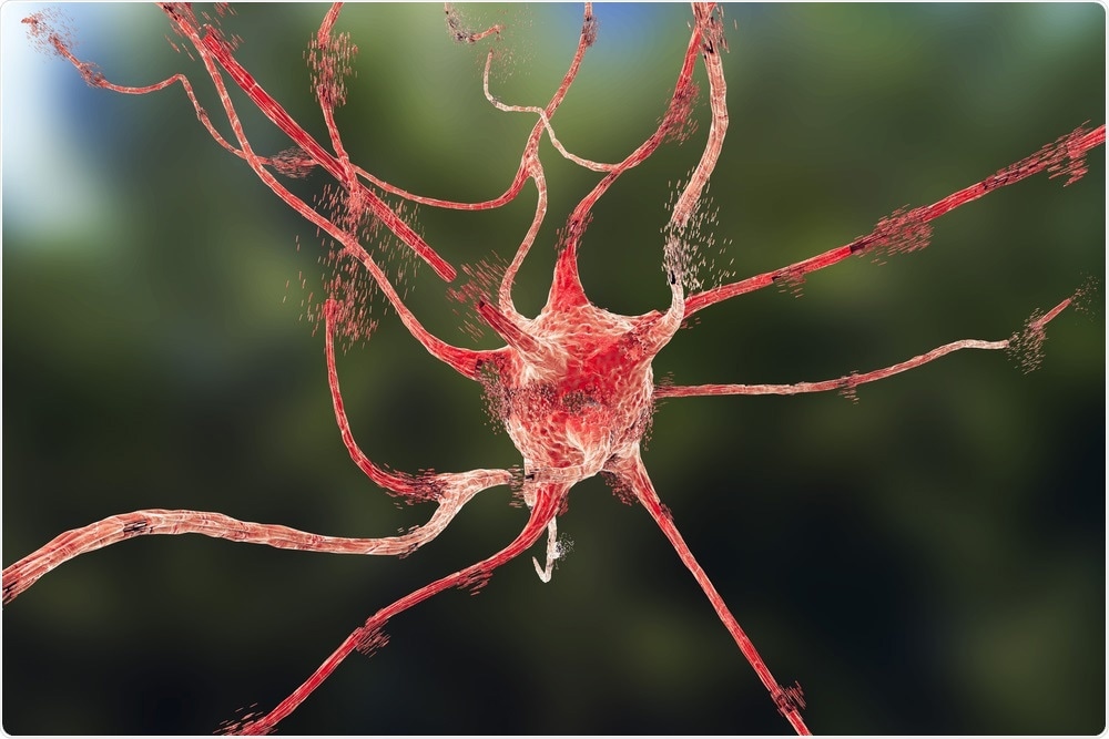
 Kateryna Kon | Shutterstock
What metabolic changes are typically seen in the injured brain?
For severe brain injuries, we usually monitor metabolites like glucose, lactate, and pyruvate, which tell us how the brain is making energy. There are specific patterns that can tell us if the brain is recovering or not.
For example, if the brain is repairing itself, it is going to consume a lot of glucose, so you can see the concentration go down. A lot of lactate and pyruvate are produced in this process, so you will see the concentrations go up. That is a good sign. On the contrary, if the brain can’t produce energy, you will see lactate go up and pyruvate go down. This can be detected with brain microdialysis, and probably with microelectrode biosensors in the future.
Why are microelectrode biosensors useful for the detection of neurochemical changes in brain interstitial fluid?
Microelectrode biosensors are useful because they can be miniaturized to very small sizes. This helps in preventing injury to the brain when inserting the probe. Another advantage is that they can provide second by second data, allowing us to observe very fast and transient changes going on in the brain.
What are the major limitations of conventional microelectrode biosensors?
The major limitation is the size of the probes. We have to insert the probe in order to understand the chemistry inside the brain. This can injure the brain, specifically the small blood vessels that are in the tissue. This creates two problems. Firstly, it causes further damage to the brain, and secondly, it reduces the validity of the data.
Even if we do not cause any damage, another problem is that we do not know if the surrounding tissue behaves differently when a probe has been inserted. However, microelectrode biosensors are currently tested in animals and represent a major advance compared to conventional techniques such as intracerebral microdialysis.
Please can you describe the ultra-small microelectrode biosensors that you developed?
Microelectrode biosensors consist of a microelectrode with an enzyme immobilized on the tip. It is basically an enzyme assay that is miniaturized to a very small size. Conventional microelectrode biosensors carry many risks, as mentioned above. Within my research team, we developed an ultra-small biosensor that is 12 microns in diameter using carbon fibers. This is incredibly small and we are proud to have achieved that.
BioSensors in Brain Trauma
BioSensors in Brain Trauma from
AZoNetwork on
Vimeo.
What did you see when the biosensors were implanted into a rat model? Why was this significant?
When the biosensors were implanted into a rat model, we saw decreased oxygen levels and lactate levels in the brain. This proved that the concentration estimates we had come up with based on data from conventional microelectrode biosensors were not completely accurate. We were then able to adjust the estimates to something that is closer to reality, so the testing was really important.
We also found that the small blood vessels around the sensor were preserved. This suggests that our probes are less likely to injure the surrounding tissue when they are inserted and removed. This will be important if we want to apply our sensors to patients.
Why did you choose to use platinized carbon fibers?
Platinum is really the material of choice for making microelectrode biosensors. However, the problem with platinum wires is that they tend to be thick. Furthermore, platinum is a soft metal, so if you make the wires too thin, the whole thing will bend, making it impossible to manipulate.
To solve this issue, we turned to carbon fibers. Carbon fibers are very rigid and can be manipulated easily. The only problem with carbon is that it is not a good electrochemical material for doing the kind of assays we want to do.
So, we decided to combine the two properties by devising a way to cover the carbon fiber with platinum. By doing this, we obtained an ultra-small object with all of the chemical properties of platinum, giving us both advantages together.
Why is it important to be able to monitor the brain second-by-second after a TBI?
We know that in the injured brain there are some very rapid and transient pathological events that we want to be able to see, so it is important for any technique to have a high temporal resolution. An example of this is cortical spreading depolarization, where waves of depolarization spread across the brain via nerves and glial cells. These cells are depolarized together and propagate into the brain before vanishing. The whole process lasts about five minutes.
We need to be able to describe each phase of processes like cortical spreading depolarization for the physicians to understand whether these waves occur, what the chemical signatures are, and if it poses a threat to the health of the patient or not. Being able to observe this second-by-second is excellent. We could also have one data point for ten seconds, but second by second is really what we were aiming for.
What other benefits do the new biosensors offer over conventional biosensors?
The main advantage of our ultra-small biosensors is their size, which allows us to implant them and take them out without injury to the patient or the animal. In addition, because we are not injuring the tissue, our chemical values are much more accurate and relevant to the real state of the brain we want to study. This, in turn, allows physicians to make the best possible clinical decisions for their patients.

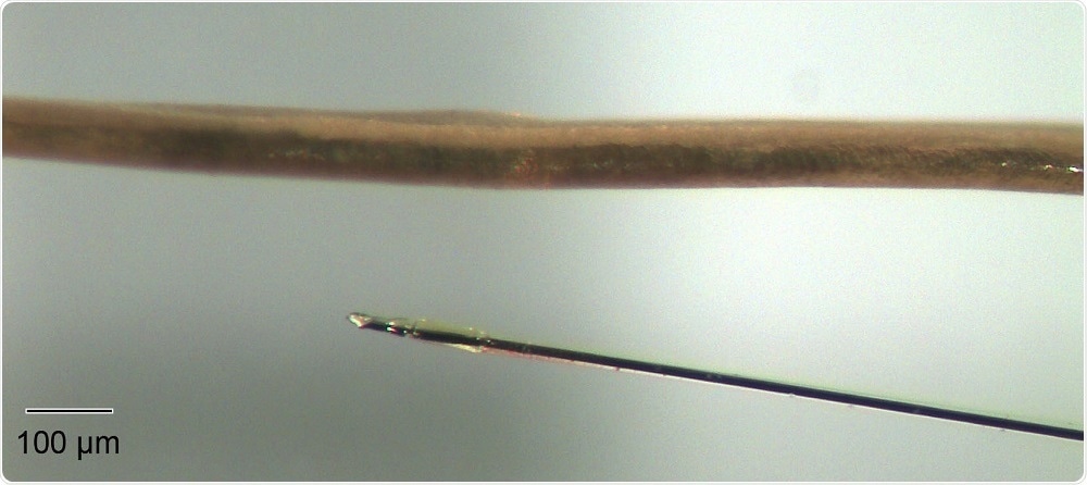
An ultra-small microelectrode biosensor next to a human hair. This image demonstrates the degree of miniaturization that Marinesco's team were able to achieve.
In the future, do you think ultra-small microelectrode biosensors will enter the clinic? What are the next steps for your research?
I really hope that the biosensors we have developed enter the clinic – this is the overall goal of my research and my life. I think they will provide a lot of advantages, given that they provide second-by-second monitoring with very little lesion to brain tissue. However, there is still a long way to go until we can start to implement them in patients…
There are several things we need to do. Firstly, we need to make sure that these devices are not toxic and that they will not break in the patient brain. That would be a disaster for the patient, so we do not want that.
We also need to make sure that they will work for seven to fourteen days in the brain. That is the length of time we need to monitor the patients, as they can have complications for up to two weeks after they are admitted to the hospital, and we are not quite there yet. There is still a long way to go, but my hope is our biosensors will one day serve the public.
Where can readers find more information?
Our latest paper on minimally invasive microelectrode biosensors based on platinized carbon fibers: Chatard C, Sabac A, Moreno-Velasquez L, Meiller A and Marinesco S (2018) Minimally invasive microelectrode biosensors for brain monitoring based on platinized carbon fibers. ACS Central Science. 4: 1751-60. doi-org.gate2.inist.fr/10.1021/acscentsci.8b00797
A recent paper on monitoring traumatic brain injury in animals using microelectrode biosensors: Balança B, Meiller A, Bezin L, Dreier J, Lieutaud T and Marinesco S (2017) Altered hypermetabolic response to cortical spreading depolarizations after traumatic brain injury in rats. J Cereb Blood Flow Metab. 37(5):1670-1686. doi.org/10.1177/0271678X16657571
Recent review on the topic: Chatard C, Meiller A and Marinesco S (2018) Microelectrode biosensors for in vivo analysis of brain interstitial fluid. Electroanalysis 30: 977-998. doi.org/10.1002/elan.201700836
About Stéphane Marinesco
 Stéphane Marinesco was trained in engineering at Ecole Polytechnique (France) and received a PhD in Neuroscience from the University of Lyon working on serotonin detection in the rat brain and studying its role in sleep and stress.
Dr. Marinesco received further training as a postdoctoral researcher at Yale University and UC Irvine with Pr Thomas J Carew, studying serotonin neuromodulation in a marine mollusk using electrochemical techniques. Dr. Marinesco returned to France in 2005 as an assistant professor in CNRS at Gif sur Yvette, and an associate professor in Lyon in 2008.
Stéphane Marinesco’s current research interests focus on developing innovative microelectrode biosensors for brain monitoring and apply them to the study of neurological injury after subarachnoid hemorrhage or traumatic brain injury. His group is based in the Lyon Neuroscience Research Center.
About Pittcon
 Pittcon® is a registered trademark of The Pittsburgh Conference on Analytical Chemistry and Applied Spectroscopy, a Pennsylvania non-profit organization. Co-sponsored by the Spectroscopy Society of Pittsburgh and the Society for Analytical Chemists of Pittsburgh, Pittcon is the premier annual conference and exposition on laboratory science.
Proceeds from Pittcon fund science education and outreach at all levels, kindergarten through adult. Pittcon donates more than a million dollars a year to provide financial and administrative support for various science outreach activities including science equipment grants, research grants, scholarships and internships for students, awards to teachers and professors, and grants to public science centers, libraries and museums.
Visit pittcon.org for more information.