Researchers at the National Institutes of Health (NIH) and Siemens have developed a high-performance, low magnetic-field MRI system that produces superior images of the body’s internal structures compared to current MRI systems. The new system may also be safer for patients with pacemakers and defibrillators.
While developments in magnetic resonance imaging have generally focused on creating systems with higher magnetic field strengths in order to produce clearer images of the brain, the NIH and Siemens researchers discovered that using the same systems with a modified magnetic field strength could produce high-quality images of the heart and lungs.
Cardiology tools that posed a heating risk when used with high-field MRI systems could now be used safely for real-time, image-guided procedures like heart catheterization as a result of the research.
Robert Balaban, Ph.D., Scientific Director of the Division of Intramural Research and chief of the Laboratory of Cardiac Energetics at NHLBI said:
We continue to explore how MRI can be optimized for diagnostic and therapeutic applications. The system reduces the risk of heating – a major barrier to the use of MRI-guided therapeutic approaches that have hampered the imaging field for decades.”
Publishing the results in Radiology, Author of the study Adrienne Campbell-Washburn, Ph.D., who is a staff scientist in the Cardiovascular Branch at NHLBI said that the results also suggest improvements could be made in producing images of the spine, brain, and abdomen. Additionally, speech and sleep disorders may benefit from this new machinery, as they will be better able to image the upper airway.
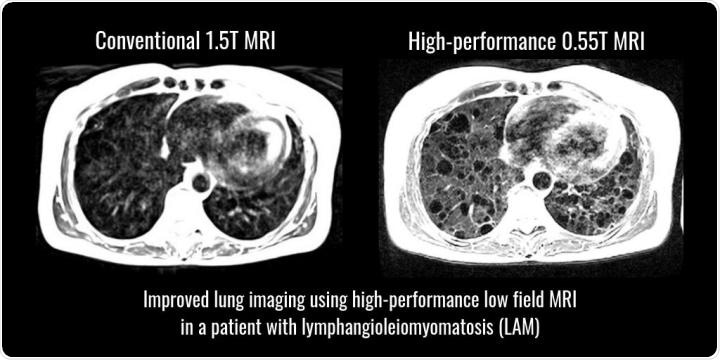
 Lung cysts and surrounding tissues in a patient with lymphangioleiomyomatosis (LAM) seen more clearly using high-performance low field MRI compared to standard MRI. Credit: Campbell-Washburn A E, et al. Opportunities in interventional and diagnostic imaging by using high-performance low-field-strength MRI. Radiological Society of North America, 2019.
“MRI of the lung is notoriously difficult and has been off-limits for years because air causes distortion in MRI images,” explained Campbell-Washburn. “A low-field MRI system equipped with contemporary imaging technology allows us to see the lungs very clearly. Plus, we can use inhaled oxygen as a contrast agent. This lets us study the structure and the function of the lungs much better.”
To achieve these improvements, the NIH and Siemens researchers modified a Siemens Healthcare MAGNETOM Aera MRI set-up, a commercially available system, to run at 0.55 tesla (T) compared to its usual rate of 1.5 T. After testing the system with objects with a consistency similar to human tissue, healthy volunteers and patients with disease undertook procedures using the new system.
Comparing the images taken at 1.5 T with images taken at 0.55 T, researchers were able to more clearly see lung cysts and surrounding tissues in images taken of patients with lymphangioleiomyomatosis (LAM), a rare, progressive condition affecting young women in which smooth muscle cells grow throughout the lungs, pulmonary blood vessels, and lymphatic system, distorting lung structure and impairing function.
Inhaled oxygen increased the brightness of the lung tissue, clearly showing advantages to using a lower-field MRI system, as patients having images taken of their lungs could be imaged without the use of a contrast agent, which poses a number of small risks.
Other advantages of low-field MRI systems is their lower operational costs as permanent scanners do not need liquid helium or specialized maintenance, and the lower fringe field means that anesthetic and monitoring equipment can be brought closer to the low-field MRI system without risk to the patient or staff.
Additionally, low-field MRI systems could be quieter than current systems, as well as being easier to maintain and initially install.
This research helps us to define new strategies that may improve accessibility and affordability of MRI as an imaging modality. We believe the high-performance, low-field MRI will have a great impact on clinical care.”
Dr. Arthur Kaindl, Head of Magnetic Resonance at Siemens Healthineers
“We can start thinking about doing more complex procedures under MRI-guidance now that we can combine standard devices with good quality cardiac imaging,” Campbell-Washburn added.
Journal reference:
Campbell-Washburn, A. E., et al. (2019). Opportunities in Interventional and Diagnostic Imaging by Using High-performance Low-Field-Strength MRI. Radiology. DOI: 10.1148/radiol.2019190452