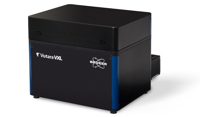
Bruker Corporation today announced the release of the Vutara VXL Super-Resolution Fluorescence Microscope for nanoscale biological imaging. The new system opens an affordable and easy-to-use path for both core facilities and individual investigators to enter the world of super-resolution imaging by incorporating Bruker’s industry-leading single-molecule localization (SML) technology in a streamlined system with compact footprint.

Bruker's Vutara VXL Super-Resolution Fluorescence Microscope.
Vutara VXL serves as a biological microscopy workstation for research on DNA, RNA and proteins, from macromolecular complexes and super-structures, to chromatin structure and chromosomal substructures, to studying functional relationships in genomes and in various subcellular organelles. This novel system also supports advanced spatial biology research in extracellular matrix structures, extracellular vesicles (EV), virology, neuroscience, and live-cell imaging. When combined with Bruker’s unique microscope fluidics unit, Vutara VXL enables multiplexed imaging for targeted, sub-micrometer multiomics in genomics, transcriptomics, and proteomics research. The Vutara VXL software already supports leading-edge methods, such as OligoSTORM, Optical Reconstruction of Chromatin Architecture (ORCA), and DNA PAINT labeling methodologies.
I am thrilled about the release of Vutara VXL, as this instrument is fundamental in making SML microscopy more accessible to the scientific community, and to the spatial 3D genomics research community in particular. Witnessing first-hand Bruker’s software and hardware development to enhance the spatial genomics capabilities that are required for my research bears testament to both their dedication and the functionality and versatility of the Vutara platform.”
Jennifer E. Phillips-Cremins, Ph.D., Associate Professor of Biotechnology and Genetics, University of Pennsylvania
We believe that enabling 3D single-molecule localization microscopy in the hands of more researchers will further advance a better understanding of cellular biology at the nano level, particularly in the emerging field of spatial omics imaging. Combined with its large field of view and ability to perform optical nanoscopy, Vutara VXL delivers multimodal capabilities and high-throughput data acquisition to enable a wider range of studies for spatially resolved genomics and transcriptomics.”
Xiaomei Li, Ph.D., Vice President and General Manager, Bruker’s Fluorescence Microscopy Business
About Vutara VXL
Based on the Vutara super-resolution platform’s proprietary biplane technology, proven industry-leading capabilities with fully integrated fluidics with unlimited multiplexing potential, and powerful analytical software, the new Vutara VXL facilitates scientific research from experiment initiation and data collection through analysis to publication. The system enables researchers to obtain intrinsic 3D super-resolution data and achieve 20 nm localization precision in XY, 50 nm in Z for organic dyes, and even finer with DNA PAINT probes.
A proprietary emission path allows the system to achieve low background fluorescence, even in thick samples, routinely imaging large Z volumes in tissue slices up to 30 μm in thickness at arbitrary distances from the coverslip. With the SRX software package and its Quantitative Localization Microscopy analysis suite, Vutara VXL facilitates the entire imaging process from acquisition and localization, and provides in-depth visual and quantitative information from biological samples in a single, seamless workflow.
Utilizing real-time localization analysis and data visualization, multi-location recording capabilities, and scripting functions for automated data analysis, completion of an entire workflow cycle can be accomplished in a matter of minutes, providing a significant enhancement to work throughput. As a multi-modality workstation for single-molecule localization microscopy as well as conventional diffraction-limited widefield imaging, Vutara VXL is an ideal platform for bioinformatics, such as looking at the spatial distribution of the transcriptome in conjunction with high-resolution genomic or proteomic imaging.