Severe acute respiratory syndrome coronavirus 2 (SARS-CoV-2), the pathogen responsible for the coronavirus disease 2019 (COVID-19) pandemic, has spread to over 58.8 million worldwide and caused over 1.3 million deaths. The rush to develop safe and effective vaccines to prevent the ongoing spread of SARS-CoV-2 is nearing the finish line as several promising candidates await regulatory body approval for widescale distribution and use.
A team of researchers from the USA and China have engineered a receptor-binding domain (RBD) that they claim improves the immunogenicity of multivalent SARS-CoV-2 vaccines. Their study, which was released on the bioRxiv* in November 2020, may help advance even more efficacious SARS-CoV-2 vaccine candidates in the future.
The spike protein versus the RBD
The spike protein of SARS-CoV-2 is key in mediating viral entry into the target host cell through its binding to the host cell receptor, the angiotensin-converting enzyme 2 (ACE2). The spike, or S, protein, binds to the receptor via the RBD, which is about 200 residues in length. It is composed of two domains, S1 and S2. The first is concerned with binding to the receptor, while the second mediates viral fusion and entry.

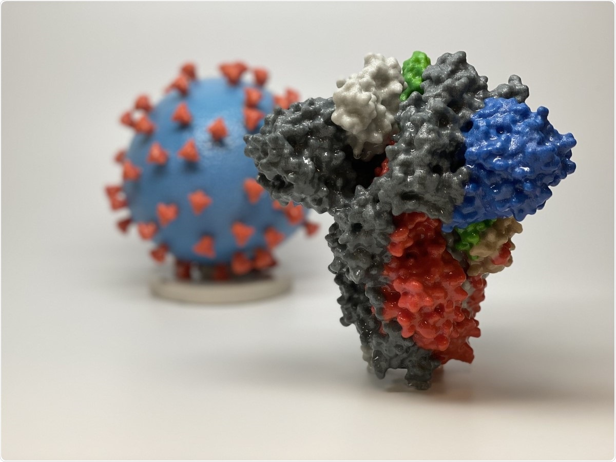
Novel Coronavirus SARS-CoV-2 Spike Protein. 3D print of a spike protein of SARS-CoV-2—also known as 2019-nCoV, the virus that causes COVID-19—in front of a 3D print of a SARS-CoV-2 virus particle. The spike protein (foreground) enables the virus to enter and infect human cells. On the virus model, the virus surface (blue) is covered with spike proteins (red) that enable the virus to enter and infect human cells. For more information, visit the NIH 3D Print Exchange at 3dprint.nih.gov. Credit: NIH. Image Credit: NIAID / Flickr

 This news article was a review of a preliminary scientific report that had not undergone peer-review at the time of publication. Since its initial publication, the scientific report has now been peer reviewed and accepted for publication in a Scientific Journal. Links to the preliminary and peer-reviewed reports are available in the Sources section at the bottom of this article. View Sources
The exposed nature of the S protein and the fact that its binding can be inhibited by specific neutralizing antibodies makes it one of the major targets for COVID-19 vaccines. It is often used in a trimeric state, after stabilization, typically by adding a pair of proline residues (S-2P).
Immunological studies show the RBD to be the primary neutralizing epitope of the virus, and as such, underscore it as a key target for any COVID-19 vaccine. While the RBD is part of the S1 subunit, it is also expressed and folded independently, with its own distinctive structure and function. Within the RBD is a variable region called the receptor-binding motif (RBM), which is the actual ACE2 binding site. The RBD has some important advantages over the S protein.
It should theoretically be easier to produce a vaccine against this virus than many others for the following reasons. Firstly, the genome is very large and is protected against mutations by an efficient proofreading system, making variations slow to emerge. In fact, the RBD has seen few mutations over the course of the current pandemic.
Secondly, the virus transmission is rapid, even before the host immune response comes into play. In fact, the RBD is exposed, despite its key role in infection of the host cell. As a result, almost all exposed RBD epitopes are neutralizing compared to a much lower proportion of epitopes on the native S protein. This means that almost all the antibodies elicited by the RBD will be neutralizing.
Thirdly, the RBD is compact and stable, allowing multiple methods of cost-effective vaccine manufacture to be applied. Viral vectors or mRNA can be used for production.
The use of RBD in a vaccine will also reduce the risk of molecular mimicry and subsequent autoimmune disease, as well as antibody-dependent enhancement of the disease, because of the fewer number of epitopes overall (as well as of linear or conformational epitopes). This will limit the possibility of side effects and allow smaller doses of the vaccine. The low proportion of decoy epitopes (responsible for binding non-neutralizing antibodies) increases its immunogenicity and neutralizing capacity.
In the current study, the researchers confirmed that the SARS-CoV-2 RBD elicits a powerful neutralizing response in the form of antibodies that prevent the S protein from binding to the ACE2 receptor. However, these antibodies do not cause ADE.
The use of carrier proteins
If multiple antigens are presented in a vaccine, by fusing the antigen to a multivalent carrier protein such as the H. pylori ferritin 24-mer protein, the immunogenicity is much higher compared to dimers or trimers. This process, again, is much easier with the small RBD than with the full-length trimeric S protein.
The researchers observed that the use of either of the two carrier proteins, conjugated to the native RBD, along with a powerful adjuvant, heightened the potency of the neutralizing responses in a rodent experiment, compared to the use of a stabilized S protein ectodomain following its conjugation to the same carriers. However, the RBD is poorly expressed by itself or in conjugation with a carrier protein, making it unsuitable as a vaccine antigen.
Glycosylation of the RBD
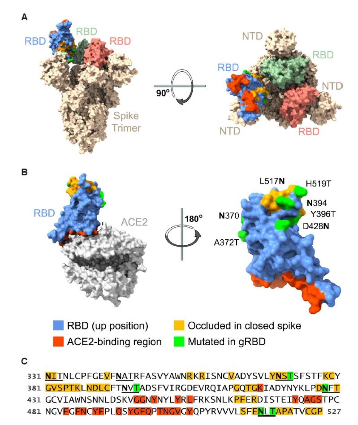
The researchers describe the modification of the native RBD by the introduction of four novel glycosylation sites. This glycosylated RBD (gRBD) was expressed far more efficiently, they found. Unexpectedly, it also showed the ability to induce a greater neutralization capacity.
This could be because the glycans improve the RBD folding or solubility, or mask immunodominant epitopes which elicit non-neutralizing antibodies.
The gRBD also acts as a highly immunogenic protein relative to the wild-type RBD when used in a protein subunit vaccine. The gRBD could be conjugated with any of five multivalent carrier proteins for expression as a DNA vaccine antigen. In this form, it was associated with still more powerful neutralization capacity, compared to either the native RBD or S-2P. The H. pylori ferritin 24-mer protein showed the capacity to elicit the most potent neutralizing antibodies, among the tested carrier proteins. This could be due to the multivalency, its higher expression and the presence of epitopes for T cells.

Engineered SARS-CoV-2 RBD glycans enhance expression of multivalent RBD fusion proteins. Views of the RBD (A) in the context of the SARS-CoV-2 S protein in the open one-up conformation, with the ACE2-binding region (red) facing upward and (B) bound to the ACE2 receptor, with the RBD ACE2-binding region facing downwards. Blue indicates surface residues that are neither occluded in the closed conformation (indicated by yellow) nor part of the ACE2 interface (red). Green indicates residues whose mutation creates a novel N-glycoslylation motif. (C) The sequence of the engineered RBD bearing four novel glycosylation motifs (gRBD) is shown. Numbering indicates S-protein residue. Glycosylation motifs (2 native and 4 engineered) are underlined. Coloring is as described in (B).
Conclusion
The researchers conclude, “Our data suggest that multivalent gRBD antigens can reduce costs and doses, and improve the immunogenicity, of all major classes of SARS-CoV-2 vaccines.”

 This news article was a review of a preliminary scientific report that had not undergone peer-review at the time of publication. Since its initial publication, the scientific report has now been peer reviewed and accepted for publication in a Scientific Journal. Links to the preliminary and peer-reviewed reports are available in the Sources section at the bottom of this article. View Sources