Coronavirus disease 2019 (COVID-19) emerged in Wuhan, China, and has since spread to nearly every country globally, causing over five million deaths and forcing many countries into costly and restrictive measures to limit transmission. COVID-19 causes a wide range of symptoms. Many infections present completely asymptomatically, but in the most severe cases, progressive lung damage is associated with the breakdown of blood vessels, edema, endothelins, thrombosis, and inflammatory cell infiltration, along with many other vascular-related pathological events.
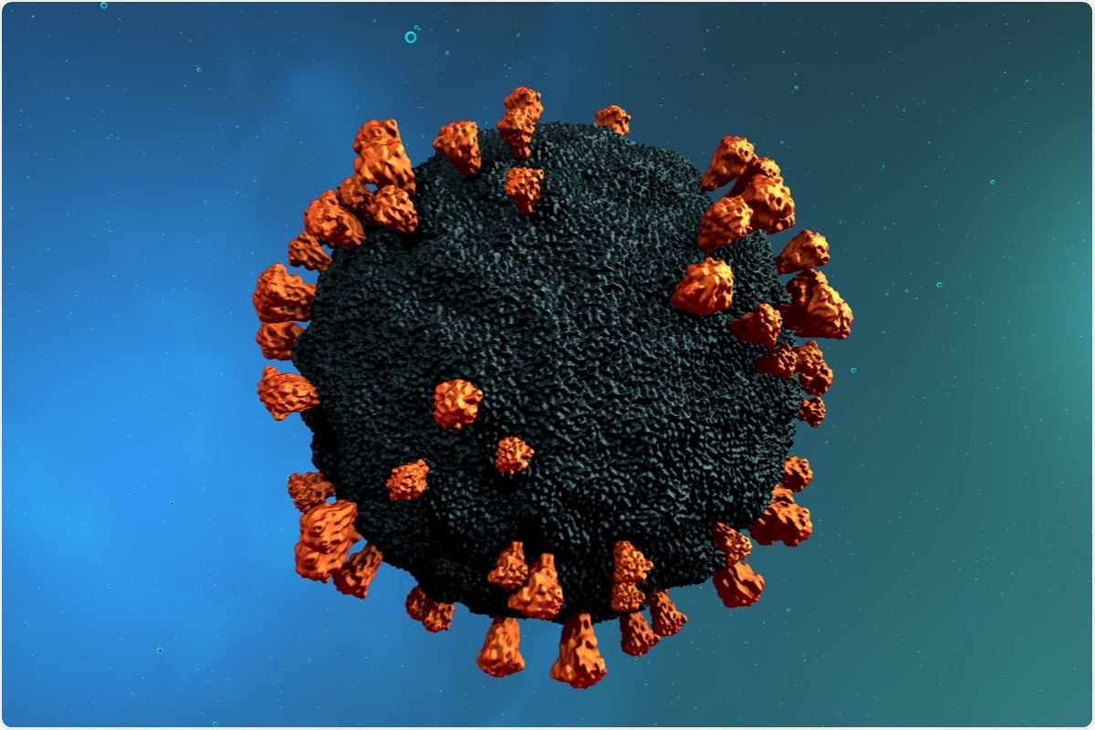
 Study: Endothelial cells are not productively infected by SARS-CoV-2. Image Credit: CROCOTHERY/ Shutterstock
It is unclear whether these events are caused by inflammation-induced endothelial activation or viral infection of the endothelium. In a paper published in Clinical & Translational Immunology, researchers from the University of Queensland have investigated whether infection of endothelial cells with severe acute respiratory syndrome coronavirus 2 (SARS-CoV-2) is possible.
Background
The spike protein of SARS-CoV-2 is essential to the disease's pathogenicity. The S1 subunit contains a receptor-binding domain (RBD) that can bind to angiotensin-converting enzyme 2 (ACE2) to permit viral cell entry, while the N-terminal domain of the S2 subunit is responsible for membrane fusion. The spike protein requires cleavage to activate. The cleavage of the spike proteins can be performed by host surface proteases such as TMPRSS2. While there are alternate routes of cleavage, this is the most common.
While endothelial cells are known to express both TMPRSS2 and ACE2, it is currently unknown whether SARS-CoV-2 can infect endothelial cells. Infection of this nature has been seen in culture but never detected in deceased COVID-19 patients, and the pathway by which SARS-CoV-2 would come into contact with endothelial cells remains unclear. It is possible that the epithelial cells in the lungs could lose their function as a barrier through infection, or rare cases of the virus spreading through the bloodstream could expose endothelial cells.
The study
To determine how prevalent endothelial cell infection with SARS-CoV-2 is in vivo, the researchers obtained autopsy lung samples from deceased COVID-19 patients. Two out of ten were positive for COVID-19 RNA in the lungs, but spike mRNA could not be found in endothelial cells.
Following this, in vitro cultures of human umbilical vein endothelial cells (HUVECs) and human microvascular endothelial lung cells (HMVEC-L) were tested for expression of ACE2 and TMPRSS2, with immunofluorescence and Western blots showing low levels of expression.
The ratio of non-glycosylated ACE2 was also lower in endothelial cells than epithelial cells – and several studies have suggested glycosylated ACE2 can resist SARS-CoV-2 spike protein binding. However, endothelial cells were confirmed to express Neuropilin-1, an alternate surface protein that the spike protein RBD can bind to and enter through.
Next, the researchers examined whether endothelial cells were susceptible to infection when apically or basolaterally exposed to the disease. Once again, no infectious virus was discovered in the supernatant of these cells anywhere up to 72 hours post-infection, and no SARS-CoV-2 nucleocapsid protein was detected by immunofluorescence or Western blot.
Following these results, higher and higher viral titers were used when exposing the endothelial cells. Some small viral titers were detected, with increased nucleocapsid protein seen in cells both infected apically and basolaterally. This shows that infection of endothelial cells is possible but abortive. When ACE2 was overexpressed in endothelial cells, active viral replication was possible, suggesting low ACE2 expression protects wild-type endothelial cells from SARS-CoV-2 infection.
When the researchers examined the infected, nucleocapsid positive endothelial cells, they discovered that they showed unusual conformations and were positive for cleaved caspase 3, strongly suggesting that infected endothelial cells rapidly enter apoptosis.
Following this, the researchers examined the immune response induced by abortive SARS-CoV-2 infection in endothelial cells. A leukocyte adhesion molecule, LCAM-1, is known to be induced when endothelial cells are inflamed. HMVEC-L cells showed increased LCAM-1 expression following SARS-CoV-2 exposure, and other inflammatory agents showed higher levels in endothelial cells following exposure.
Conclusion
The authors have proved that effective infection of endothelial cells by SARS-CoV-2 is all but impossible in regular infection. The activation of inflammatory signaling pathways likely causes the endothelial disruption seen in patients with severe SARS-CoV-2.
This information could prove invaluable for health care workers and those attempting to repurpose/ discover drugs effective against severe disease in COVID-19 patients, as it could help them to decide targets to focus on and avoid research into routes that would not work, such as attempting to prevent infection of endothelial cells. The researchers suggest targeting the inflammatory response is the best option to prevent endothelial dysfunction.
Journal reference:
Schimmel, L., et al. (2021), Endothelial cells are not productively infected by SARS-CoV-2. Clin Transl Immunol. doi: doi.org/10.1002/cti2.1350