It aimed to investigate whether hyperglycemia persisted in these patients during the six-month follow-up and its possible causes. Specifically, they evaluated insulin resistance (IR) and beta cell dysfunction as possible causes of COVID-19-induced hyperglycemia.

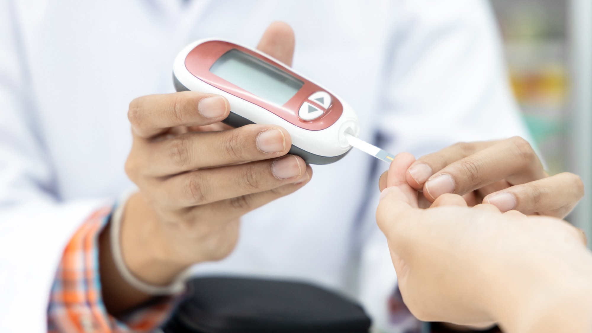
Study: Severe COVID-19 associated hyperglycemia is caused by beta cell dysfunction: a prospective cohort study. Image Credit: Darunrat Wongsuvan/Shutterstock.com
Background
The prevalence of new-onset diabetes and hyperglycemia was unexpectedly high during the acute stage of COVID-19. Research suggests that insulin resistance (IR) and beta cell dysfunction resulted in these fatal metabolic disturbances in patients with acute COVID-19.
IR decreases skeletal muscle and liver insulin sensitivity as a stress response because catecholamines and cortisol demand are high during the acute phase of COVID-19. It also increases lipolysis and elevates circulating non-esterified fatty acids (NEFA).
Severe acute respiratory syndrome coronavirus 2 (SARS-CoV-2)-triggered inflammation increases the energy demand in affected patients, accompanied by increased reactive oxygen species (ROS) production.
It activates the renin-angiotensin-aldosterone system (RAAS) in pancreatic beta cells. Some studies suggest that SARS-CoV-2 replication directly damages beta cells. Though insulin-glycemic indices tended to improve, hyperglycemia persisted in these patients over time, up to two months in some cases.
About the study
In the present study, researchers screened patients (aged over 18 years) at specialized COVID-19 wards of the University Hospital Královské Vinohrady in Prague during March–April 2021, as soon as oxygen support was removed, i.e., baseline (T0).
It helped them capture the acute phase of illness when they suffered from COVID-19-induced bilateral pneumonia. The team enrolled 37 patients based on the eligibility criteria and re-examined them after three months (T3) and six months (T6), respectively. Specifically, they compared patients with normal glycemia vs. hyperglycemia at T0 and T6.
The team performed anthropometric measurements and medical examinations besides collecting each participant's blood samples for the analysis of glucose homeostasis parameters, lipid profile, etc.
Additionally, they performed an oral glucose tolerance test (OGTT) per standard WHO recommendations, where an insulinogenic index (IGI) indicated insulin secretion levels. Further, they calculated the oral disposition index (DI) to determine the beta cell function of all participants.
Finally, the team computed resting energy expenditure (REE) and respiratory quotient (RQ) from the gas exchange and nitrogen losses using the Weir formula. The RQ and REE changes from baseline to 120 min OGTT served as parameters of metabolic flexibility and diet-induced thermogenesis, respectively.
Results
Data of only 26 participants with age ~59 years; 35% of women were available for the analysis at follow-up completed 21 ± 6.5 days after COVID-19 diagnosis. Of these, 19 and 18 patients needed high or low-flow nasal oxygen therapy. During hospitalization, 23, 9, and 1 patient(s) received high-dose dexamethasone, prednisone, and methyl-prednisolone treatment, respectively.
The prevalence of hyperglycemia at baseline was 65% in the study population, and it remained as high as 50% up to T6, of which 10 developed prediabetes, while three needed pharmacological treatment for type 2 diabetes. The researchers noted baseline glycated hemoglobin (HbA1c) levels >48 mmol/mol in the hyperglycemic group comprising seven patients.
In the hyperglycemia group, insulinogenic response declined and became comparable with the normoglycemic group at T6. Also, the former had worse insulinogenic and DIs at baseline; however, the difference became insignificant at T6. ISI Matsuda and HOMA indices indicating insulin sensitivity changes improved only in the hyperglycemic group by T6.
Furthermore, the total body weight of people in the hyperglycemia group increased by 3.6 ± 3.2 kg over six months. Also, while patients were hypermetabolic at T0, with REE of 30.3 ± 4.0 kcal per kg of active tissue mass, nearly 19% above the predicted values from the Harris-Benedict equation at baseline, REE declined over six months and attained the predicted values over three months.
Conclusions
Although uncertainties surround the link between COVID-19 and diabetes, the present study results clarified that these two are intertwined and interact closely. Diabetes is a risk factor for severe COVID-19, and acute COVID-19 might also induce diabetes in individuals with impaired beta-cell function.
Furthermore, study results showed that corticosteroid treatment quickly normalized hypermetabolism associated with severe COVID-19, though this treatment should last for the shortest time possible. Further research is needed to explore the mechanisms of beta cell dysfunction in COVID-19.