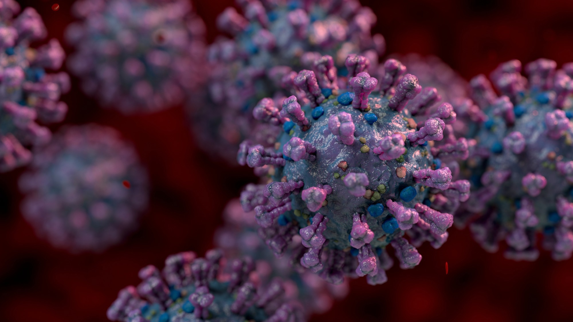
Several studies have shown that severe acute respiratory syndrome coronavirus-2 (SARS-CoV-2), the causal agent of the coronavirus disease 2019 (COVID-19) pandemic, affects multiple systems.
 Study: SARS-CoV-2 infection causes dopaminergic neuron senescence. Image Credit: Ninc Vienna/Shutterstock.com
Background
Individuals with SARS-CoV-2 infection commonly develop various neurological conditions such as headaches, anosmia and dysgeusia.
Other abnormal neurological symptoms found in these patients are seizures, acute inflammatory polyradiculoneuropathy (Guillain-Barre syndrome) and stroke. Patients with SARS-CoV-2 infection are also at a higher risk of developing psychiatric disorders.
Human pluripotent stem cells (hPSCs)-derived organoid models have revealed that choroid plexus cells of the central nervous system (CNS) are highly susceptible to SARS-CoV-2 infection. Despite this information, the tropism of SARS-CoV-2 for neurons has not been confirmed.
The hPSCs-derived organoid/cell-based platform was previously utilized to study the tropism of SARS-CoV-2. This platform revealed the association of hPSC-derived midbrain dopamine (DA) neurons (primary neurodegeneration targets in Parkinson’s disease [PD]) with SARS-CoV-2 infection.
A similar experimental protocol indicated that hPSC-derived cortical neurons were not permissive to SARS-CoV-2 infection. This finding suggested that all neurons are not unilaterally permissive to SARS-CoV-2 infection.
About the study
The current study investigated the DA neurons’ response to COVID-19. The molecular changes triggered by SARS-CoV-2 infection were also assessed.
DA neurons were differentiated from hPSCs using a previously published protocol. After studying NURR1-GFP+ cells at day 25 of the differentiation, it was confirmed that SARS-CoV-2 can infect DA neurons.
Immunostaining, scRNAseq, and real-time quantitative PCR were conducted to verify that the majority of cells in the experiment were DA neurons. Therefore, senescence phenotypes were primarily attributed to DA neurons.
scRNA-seq analysis revealed a high expression of LMO3 and an absence of CALB1 expression, suggesting that SARS-CoV-2 infects DA neurons. It must be noted that DA neurons with an A9-like subtype were more susceptible to SARS-CoV-2. A similar observation was noted in previous studies where A9 neurons were found to be vulnerable in the substantia nigra, which is most affected in PD.
Taken together, this study indicates that SARS-CoV-2 infection activates cellular senescence in DA neurons, which could contribute to PD pathogenesis. Since lethargy and anhedonia are two symptoms linked with long COVID, more research is required to understand whether they are linked with DA neuron dysfunction due to SARS-CoV-2 infection.
The response of PD-iPSC-derived DA neurons to SARS-CoV-2 infection was monitored. SNCA is a gene associated with familial PD, which encodes the protein a-synuclein (α-Syn). Previous studies have identified copy-number variations (CNVs) in SNCA in PD patients. This study used isogenic SNCA 4 copy, 2 copy, and 0 copy iPSC-derived DA neurons to assess their response to COVID-19.
In comparison to the studied SNCA copy number, SNCA 4 copy iPSC-derived DA neurons exhibited the most elevated SARS-CoV-2 infection. This occurrence could be linked to an elevated TLR4 expression that facilitates viral invasion of host cells.
The immunostaining technique also revealed that SNCA 4 copy iPSC-derived DA neurons exhibited the highest α-Syn expression compared to other SNCA copy numbers.
Future studies must focus on the underlying mechanisms that entail increased susceptibility to SARS-CoV-2 infection in SNCA 4 copy DA neurons. Interestingly, SARS-CoV-2 infection enhanced the levels of b-gal+ cells and lysosomal accumulation in all iPSC-derived DA neurons.
Previous studies have shown that α-Syn has a high affinity to the Nucleocapsid (N) protein of SARS-CoV-2. Western Blotting technique also confirmed an increased α-Syn expression post-SARS-CoV-2 infection.
Riluzole, imatinib, and metformin are FDA-approved drugs that block SARS-CoV-2-mediated DA neuron senescence, thereby, inhibiting viral infection. Imatinib was found to inhibit the viral entry in hPSC-derived lung organoid-based screen; however, a similar mode of action was not observed in riluzole.
Consistent with the findings of previous studies, the current study indicated that metformin can induce the AMPK signaling pathway in DA neurons. Scientists suspect that more unknown molecular mechanisms could be present that account for the antiviral effect of the aforementioned three drugs.
Conclusions
DA neurons were found to be the possible target for SARS-CoV-2 infection, which may activate cellular and inflammatory senescence response.
Besides DA neurons, Other cell types that include microglia and astrocytes, or pathological changes linked to a hypoxic state, could also manifest inflammatory and senescence signatures in the substantia nigra samples.
Considering the study findings, it is imperative to continually monitor COVID-19 patients to understand if they are at a higher risk of developing PD-like conditions.