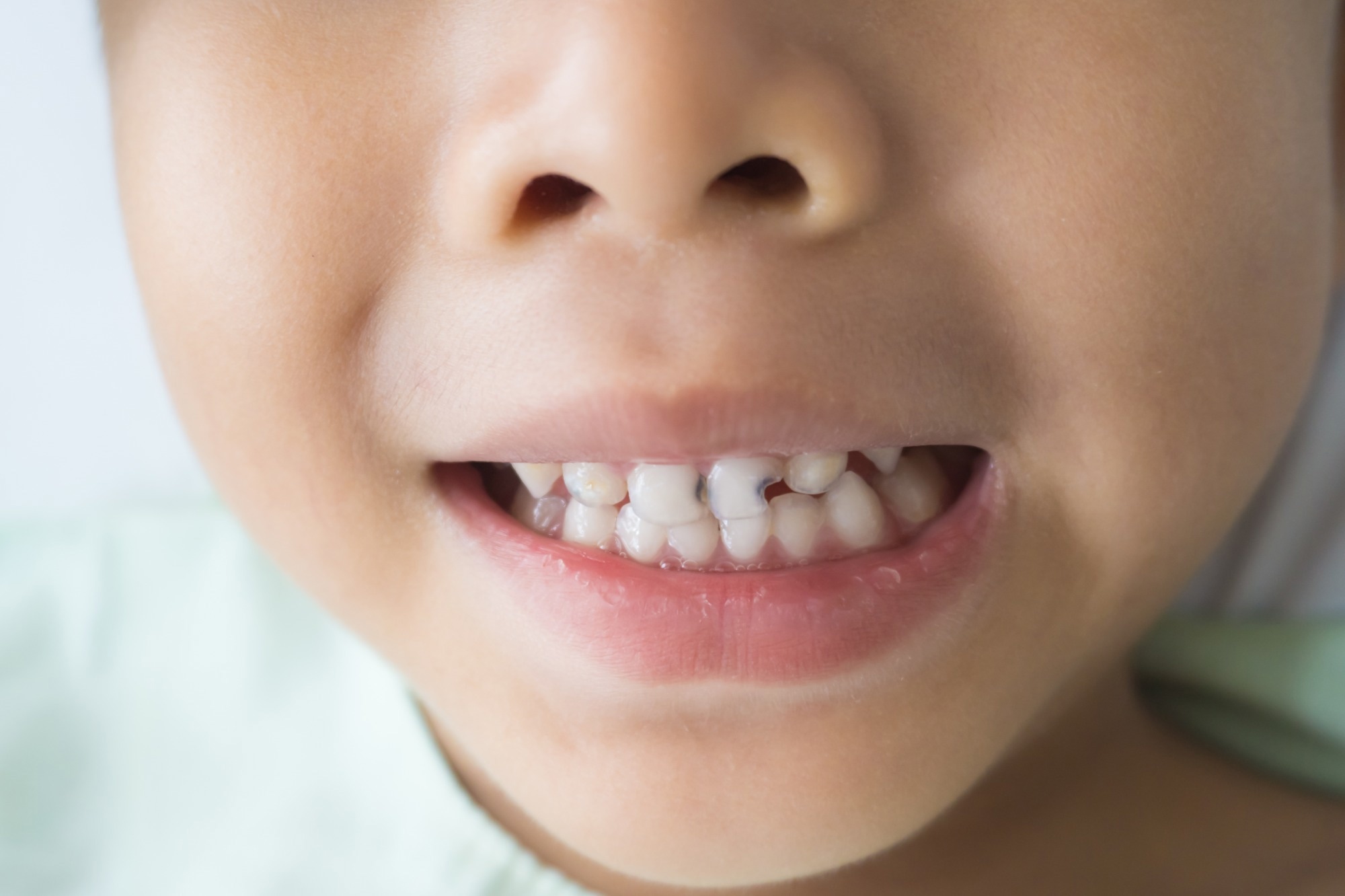
They did not find a significant association between vitamin D status and the prevalence and number of teeth affected by caries and MIH among the participants.
 Study: The association between serum vitamin D status and dental caries or molar incisor hypomineralisation in 7–9-year-old Norwegian children: a cross-sectional study. Image Credit: modina/Shutterstock.com
Background
Vitamin D, a nutrient crucial for calcium absorption and bone mineralization, may play a role in oral health through tooth development and immune modulation.
While studies on vitamin D and childhood dental caries have yielded varied results, a recent meta-analysis suggested an association between vitamin D deficiency and dental caries risk. Further, studies exploring vitamin D's impact on MIH, an enamel developmental defect, are scarce.
Previous studies have dichotomized oral health outcomes, leading to a loss of information about their distribution, and only a few investigations have applied continuous variables in regression analyses. Additionally, in prior studies, varied methods for measuring serum 25-hydroxyvitamin D (25(OH)D) may affect result in consistency.
Liquid chromatography with tandem mass spectrometry (LC-MS/MS) is considered superior to immunoassays for accurate 25(OH)D measurements.
Understanding vitamin D's influence on oral health is particularly relevant in far-north regions like Norway, where sunlight exposure is limited. A recent Norwegian study linked maternal vitamin D levels during pregnancy to MIH in offspring, but broader investigations into vitamin D's oral health impact in Norwegian children are lacking.
Therefore, the present study aimed to explore associations between LC-MS/MS-measured serum vitamin D levels and dental caries or MIH prevalence and severity in Norwegian children.
About the study
In the present cross-sectional study, data was obtained from the TRIP-study (short for TRaining In Pregnancy study), a previous randomized controlled trial measuring the impact of a pregnancy exercise program on gestational diabetes.
In a follow-up of the study, 7–9-year-old children of the recruited women were examined for their oral (TRIP-tann sub-study) and bone health (TRIP-bein sub-study). A total of 101 children with a mean age of 8.1 years were included in the study, 53% of whom were female.
Serum 25(OH)D was analyzed continuously and categorically using LC-MS/MS to indicate vitamin D intake and exposure to ultraviolet light. Parathyroid hormone (PTH) levels, indicating the functioning of calcium metabolism, were analyzed using an immunoluminometric assay (ILMA).
The oral examination, conducted by two calibrated dentists, involved the assessment of caries using the 5-grade dmft/DMFT (short for decayed, missing, and filled teeth) index. Additionally, MIH was recorded as per the MIH index and considered defects ≥1 mm on erupted teeth.
Potential confounding factors, including age, sex, body mass index (BMI), season of blood draw, and mother's education, were obtained from the study records and birth records.
Statistical analysis involved the use of odds ratios (OR), rate ratios (RR), and hurdle models incorporating logistic and negative binomial regression to evaluate the potential link between child vitamin D status and outcomes of oral health.
Results and discussion
Although the mean PTH levels were found to be normal among all children, 27% of them showed insufficient vitamin D levels. Vitamin D levels were particularly low among girls whose blood was drawn in winter or spring and whose mothers were relatively least educated.
While the prevalence of dental caries was found to be 25% among the participants, that of MIH was found to be 32%. The prevalence of dentine caries was found to be 15%, and that of yellow/brown opacities on MIH teeth was 8%. Children with insufficient vitamin D showed higher proportions of caries (+11.7%), MIH (+8.4%), mean number of teeth affected by MIH (+0.4), and mean caries experience (+0.3).
Children with insufficient vitamin D showed higher odds for caries and MIH prevalence (as compared to those with sufficient vitamin D), but without statistical significance.
The continuous analysis showed that caries risk tended to increase with decreasing vitamin D levels. The MIH analysis indicated fewer affected teeth in those with insufficient vitamin D, but the results were not statistically significant.
The researchers suggested that the lack of significant associations in the study could be potentially explained by the low caries prevalence and caries experience among the studied children.
The study's limitations include its cross-sectional design that precludes the establishment of temporality among the variables, as well as its small sample size, potential selection bias, limited statistical power for associations, inability to explore potential inverse relationships, and a lack of generalizability to the Norwegian pediatric population.
Conclusion
In summary, the prevalence and severity of dental caries and MIH among 7–9-year-old children in Norway were not found to be significantly associated with vitamin D status.
The findings warrant larger prospective studies, incorporating multiple serum vitamin D measurements and oral examinations throughout childhood, to investigate this relationship further.