CAD is a leading cause of death with a significant global burden. Accurate CAD assessment is crucial for care and treatment. Currently, pretest probability tools (PTPs) are used to determine the probability of CAD in suspected patients. Nevertheless, these tools have subjectivity issues, limited generalizability, and modest precision.
Although supplementary cardiovascular examinations (coronary artery calcium score and electrocardiography) or complex clinical models integrating additional laboratory markers and risk factors could improve probability estimates, challenges related to time efficiency, procedural complexity, and limited availability exist.
IRT, a non-contact surface temperature detection technology, has been promising for disease assessment. It can identify inflammation and abnormal blood circulation from skin temperature patterns. Studies indicate associations between IRT information and atherosclerotic cardiovascular disease and related conditions.
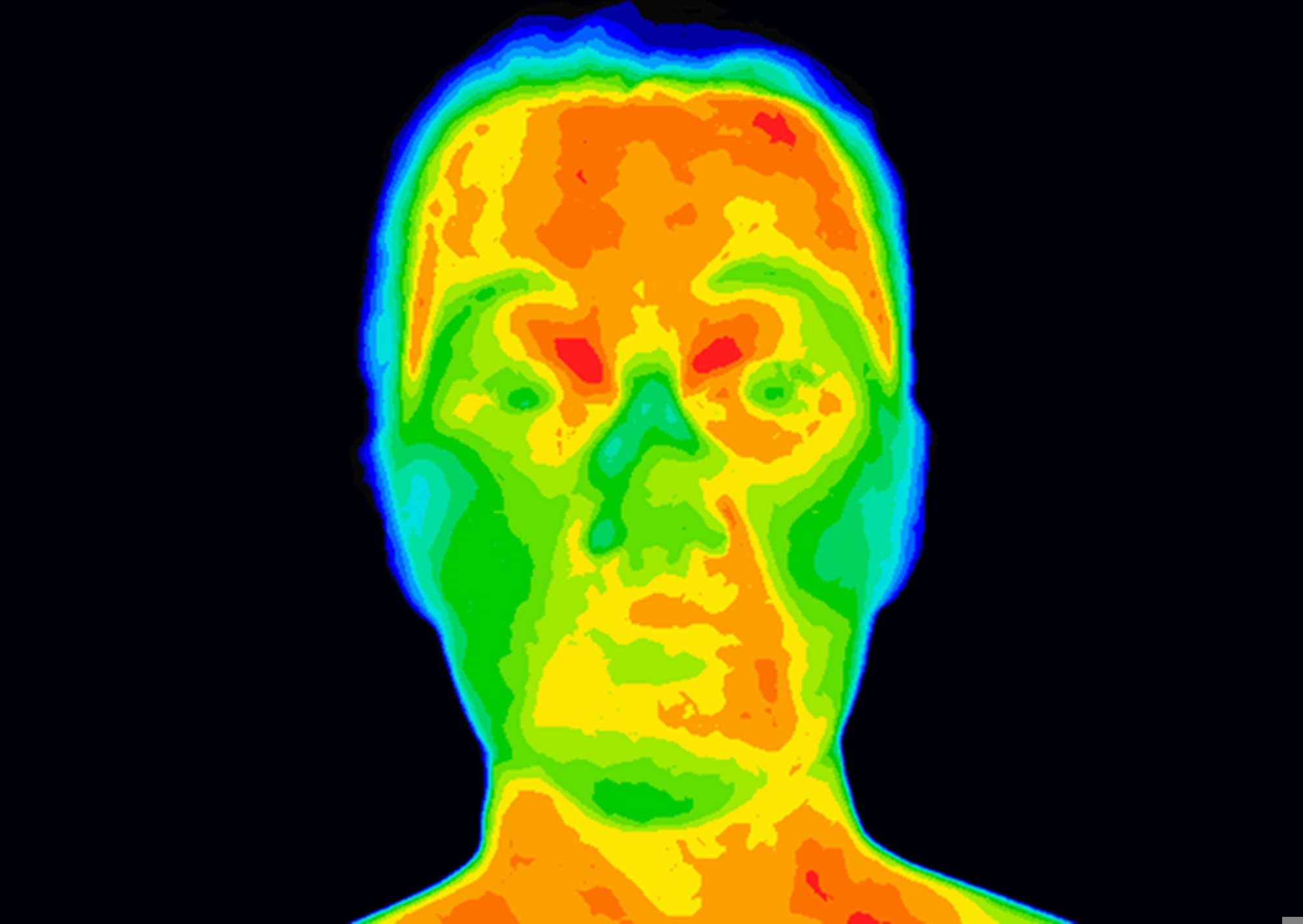
 Study: Prediction of coronary artery disease based on facial temperature information captured by non-contact infrared thermography. Image Credit: Anita van den Broek / Shutterstock
About the study
In the present study, researchers evaluated the feasibility of facial IRT temperature data for CAD prediction. Adults undergoing coronary CT angiography (CCTA) or invasive coronary angiography (ICA) were enrolled. Trained personnel obtained baseline data and performed IRT filming before CCTA or ICA.
Electronic medical records were used for additional information, including blood biochemistry, clinical history, risk factors, and CAD workup findings. One IRT image was selected per participant and processed (uniform resizing, greyscale conversion, and background cropping) before analyses. The prediction of interest was the presence of CAD, defined as a coronary lesion stenosis ≥ 50%.
The team developed an IRT image model with an advanced deep-learning algorithm. Two models were also developed for comparison; one was the PTP model (the clinical baseline) that included patients’ age, sex, and symptom characteristics, while the other was a hybrid incorporating both IRT and clinical information from the IRT image and PTP models, respectively.
Several interpretation analyses were performed, including occlusion experiments, saliency map visualization, dose-response analyses, and CAD surrogate label prediction. Further, diverse IRT tabular features were extracted from the IRT image and classified into whole-face and region of interest (ROI)-specific levels.
Overall, extracted features were categorized into first-order texture, second-order texture, temperature, and fractal analysis features, respectively. The XGBoost algorithm integrated these extracted features and evaluated their predictive value for CAD. The researchers assessed performance by using all features and only temperature features.
Findings
In total, 893 adults undergoing CCTA or ICA were screened between September 2021 and February 2023. Of these, 460 participants aged 58.4, on average, were included; 27.4% were females, and 70% had CAD. CAD subjects were older and male and had a higher prevalence of risk factors compared to non-CAD individuals. The IRT image model performed substantially better than the PTP model.
However, the performance of hybrid and IRT image models was not significantly different. Using only temperature features or all extracted features had superior prediction performance, which was in line with the IRT image model. At the whole-face level, the overall left-right temperature difference had the highest impact, whereas, at the ROI-specific level, the average temperature of the left jaw had the most impact.
Varying levels of performance reduction were observed for the IRT image model when occluding different ROIs. The occlusion of the upper and lower lips region had the most significant impact. Besides, the IRT image model performed well in predicting CAD-associated surrogate labels, such as hyperlipidemia, smoking, body mass index, glycated hemoglobin, and inflammation.
Conclusions
The study illustrated the feasibility of using human facial IRT temperature data for CAD prediction. The IRT image model performed better than the guideline-recommended PTP model, highlighting its potential in CAD assessment. Further, incorporating clinical information in the IRT image model had no additional improvements, suggesting that the extracted facial IRT information already encompassed relevant CAD-related information.
Moreover, the predictive value of the IRT model was validated using interpretable IRT tabular features, which were relatively consistent with the IRT image model. Furthermore, these human-interpretable IRT features also provided insights into aspects critical for CAD prediction, such as facial temperature symmetry and distribution non-uniformity. Further studies with larger sample sizes and diverse populations are required for validation.
Journal reference:
- Kung M, Zeng J, Lin S, et al. Prediction of coronary artery disease based on facial temperature information captured by non-contact infrared thermography. BMJ Health Care Inform, 2024, DOI: 10.1136/bmjhci-2023-100942, https://informatics.bmj.com/content/31/1/e100942