Background
Cellular senescence is crucial in processes like aging, cancer, and disease. Induced by stress, it involves cell-cycle arrest, cyclin-dependent kinase (CDK) inhibitors, and a senescence-associated secretory phenotype (SASP). Senescent cells have both harmful roles, such as promoting aging and disease, and beneficial ones, like tumor suppression and tissue repair. Further research is needed to clarify their cell-type-specific roles and develop therapies that balance their positive and negative effects.
About the study
Genomic polymerase chain reaction (PCR) was performed by lysing mouse tails or tissues with a lysis buffer and proteinase K at 55°C for over six hours. The mixture was then centrifuged to collect the supernatant, which was precipitated using isopropanol to obtain genomic DNA. This DNA was washed with 70% ethanol and dissolved in deionized water. Mice were genotyped using primers specific to the knockin sequence.
For mouse embryonic fibroblast experiments, Embryonic day 13.5 (E13.5) embryos were trypsinized, and cells were collected through a cell strainer. Cells were cultured in Dulbecco's Modified Eagle Medium (DMEM) with 10% fetal bovine serum (FBS) and antibiotics, with media changes every 2-3 days. The carbon tetrachloride (CCl4)-induced liver fibrosis model involved administering CCl4 intraperitoneally in increasing doses, followed by liver collection for analysis. Only female mice were used in this model to avoid gender differences.
Nonalcoholic steatohepatitis (NASH) was induced using a choline-deficient L-amino acid diet for 12 weeks, with only male mice utilized to prevent gender-related variation. For small interfering Ribonucleic Acid (siRNA) transfection, cells were transfected with siRNA using Lipofectamine RNA interference MAX (RNAiMAX) reagent, followed by incubation in a complete medium containing FBS before harvesting for analysis.
Study results
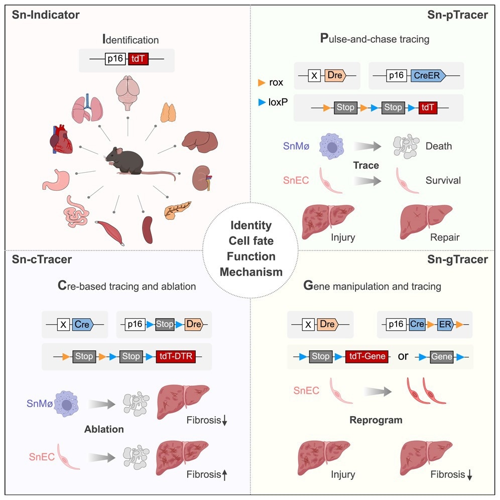
p16Ink4a+, encoded by CDK 2a (Cdkn2a), is a widely recognized marker of cellular senescence, crucial in processes like aging and disease. A fluorescence-based reporter line called p16-tdTomato (tdT) was developed to investigate its expression in different cell types, enabling single-cell resolution of p16Ink4a+ expression. Validation through mouse embryonic fibroblasts (MEFs) confirmed the senescence phenotype in p16Ink4a+ cells as they aged, characterized by increased Senescence-Associated β-Galactosidase (SA-β-Gal) staining and reduced Ki67 expression. Additionally, p16-tdT cells expressed high levels of other senescence markers like p21 and Interleukin (IL)-6, reinforcing the reliability of this model to study p16Ink4a+ expression.
Fluorescence imaging of various organs from aging p16-tdT mice revealed an increase in tdT+ cells with age, particularly in neurons of the cerebral cortex and endocrine cells in the pancreas. These tdT+ cells showed strong senescence-associated phenotypes, such as elevated SASP factors and reduced proliferative capacity, confirming the diverse senescent cell populations in aged tissues.
To investigate the roles of senescent cells in liver fibrosis, p16-tdT mice were treated with CCl4, inducing fibrosis. Enrichment of tdT+ senescent cells was observed in fibrotic areas, particularly in macrophages and endothelial cells (ECs), but not in hepatocytes. Flow cytometry and immunostaining further validated that subsets of these cell types expressed p16Ink4a+ and exhibited senescence characteristics. Single-cell RNA sequencing (scRNA-seq) corroborated these findings, showing the upregulation of senescence markers and SASP factors inp16Ink4a+ macrophages and ECs.
To trace the fate of p16Ink4a+ cells during liver injury and repair, the Sn-pTracer system was developed, enabling specific labeling of senescent macrophages and ECs. Results revealed that p16Ink4a+ macrophages, marked during injury, were depleted during repair, while ECs persisted and exhibited reduced senescence markers over time.
In addition, a new genetic tool called Sn-cTracer was developed for the ablation of p16Ink4a+ cells. Ablation of p16Ink4a+ macrophages in liver fibrosis significantly reduced fibrosis, whereas eliminating p16Ink4a+ ECs exacerbated liver injury. This differential outcome demonstrated that senescent macrophages promote fibrosis, while senescent ECs play a reparative role.
Lastly, reprogramming p16Ink4a+ ECs through Kdr overexpression in a liver injury model significantly reduced fibrosis, highlighting the therapeutic potential of targeting senescent cell populations in a cell-type-specific manner to improve tissue repair and limit fibrosis.
Novel Genetic Tools
The study introduced two powerful genetic tools, Sn-pTracer and Sn-cTracer, designed to trace and manipulate p16Ink4a+ senescent cells in specific cell types. Sn-pTracer allows for precise lineage tracing, while Sn-cTracer enables cell-type-specific ablation, overcoming the limitations of earlier models that lacked specificity.
Conclusions
To summarize, previous studies using systemic clearance of p16Ink4a+ cells lacked cell-type specificity, overlooking differences in cell identity across tissues. To address this, four genetic strategies were developed to study specific p16Ink4a+ cell types. In liver fibrosis, senescent macrophages promoted fibrosis, while senescent ECs aided in tissue repair. Ablating p16Ink4a+ macrophages reduced fibrosis, whereas eliminating p16Ink4a+ ECs impaired repair. Reprogramming ECs with Kdr overexpression enhanced their function and reduced fibrosis, offering important insights for the development of future senolytic therapies.