Breast cancer is the leading cancer affecting women globally. In 2013, the number of new cases and deaths in the US alone was 232,340 and 39,620 respectively. Treatment usually involves full or partial surgical removal of the breast, followed by chemotherapy and/or radiotherapy. Although early detection techniques have improved recently, a large proportion of women are still diagnosed in the later stages of disease, with locally advanced breast cancer (LABC), meaning their chances of long-term survival are reduced.
Neo-Adjuvant Therapy and Other Techniques
For patients with LABC, more aggressive neo-adjuvant therapy is administered for the weeks and months prior to surgery. Although a response to neo-adjuvant therapy is associated with an improved chance of survival, there is uncertainty and therefore controversy over which treatment should be chosen and how long it should be used for. The sooner oncologists can see a response to neo-adjuvant therapy, the sooner an alternative therapy can be introduced if necessary, to improve patients’ chances of survival.
The early detection of treatment responsiveness and the tailoring of treatment to individuals are therefore currently two key areas of breast cancer research. Some promising new imaging techniques have been developed to improve the detection of early responses to therapy. Traditionally, this treatment response has been assessed anatomically using physical examination, ultrasound and mammography. However, tumors can take months to shrink, even in cases where patients have responded well to their therapy. On the other hand, the ability to see a tumor’s physiology would enable oncologists to detect a patient’s response to treatment much sooner, possibly within as little as one week. Therefore, non-invasive techniques including ultrasound elastography, quantitative ultrasound and diffuse optical spectroscopy (DOS) are being demonstrated as imaging modalities that can indicate early responses to therapy.
However, researchers need to validate these modalities and show that they are of value in cancer care. To do this, researchers need to prove that clinical and pathological responses can be predicted using these techniques. Therefore, when studies of quantitative ultrasound, DOS and elastography techniques are carried out, breast tissue samples are mounted on slides after surgery for a pathologist to analyze.
Histopathology and Breast Cancer
As well as providing the basis of disease prognosis and treatment, histopathology is also the “gold standard” for validating new imaging modalities. However, beyond the traditional standard, whole-mount histopathology offers various benefits over small-slide, selective scanning. Huron Digital Pathology provides a whole slide scanning solution for studying breast cancer with improved efficiency, clarity and flexibility in the TissueScope™ scanner.

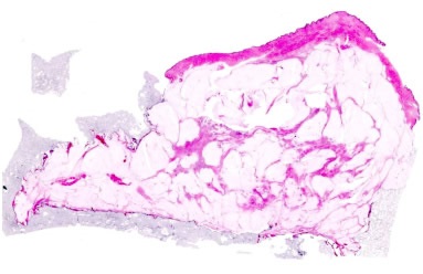
Figure 1. Image of whole-mount breast cancer specimen. Image credit: Huron Digital Pathology.
Previously, the use of small-format microscopy had meant pathologists could only analyze small, isolated breast tissue samples. However, breast specimens are relatively large, often measuring up to 11” x 11” x 3”. The pathologist’s task of determining the extent of the tumor is therefore made somewhat more difficult when only samples of 0.8” x 1.2” x .2” can be selected. The pathologist tries to describe the tumor’s extent overall, in 3D, but only a limited representation of 2D images are actually available, meaning the specimen may be undersampled and the extent of the tumor underestimated.
Using small-format histopathology, pathologists can only provide classification-level, feature-based inferences between radiographic signs and histopathologic findings.
Huron Digital Pathology’s TissueScope™
By contrast, the Huron Digital Pathology’s TissueScope™ enables large-area slides of up to 5” x 7”cm to be imaged, therefore allowing the scanning of complete mastectomies. Whole-mount histopathology using the TissueScope leads to more precise tumor measurements, with areas being captured that would otherwise be missed using traditional techniques.
Breast cancer researchers from Sweden, Canada, the US and Italy are already benefiting from the superior visual information enabled by the TissueScope in large-format histology.
The whole-mount imaging offered by TissueScope correlates with in vivo imaging, thereby providing researchers with an advantage. Aside from traditional microscopy meaning pathologists have to construct their understanding of the breast from only small samples, the breast tissue deforms easily once excised because it is high in fat content, which further obscures its shape. Since techniques such as DOS, MRI, ultrasound elastography and quantitative ultrasound can be used to image the entire breast in vivo, whole-mount histopathology provides an improved frame of reference.

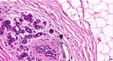
Figure 2. 20X magnification of breast tissue specimen with 0.5μm resolution. Image credit: Huron Digital Pathology.
Conclusions
Researchers are making use of the TissueScope for developing 3D images of whole specimens from individual slides, to achieve a more thorough understanding of breast tissue. Overall, new imaging modalities can be more effectively validated using the Huron Digital Pathology’s TissueScope. It offers the sharp and clear imaging quality needed for detecting tumor heterogeneity, with up to 0.25μm/pixel resolution at 40x magnification. The instrument is unmatched in the versatility and efficiency it offers because it can simultaneously scan up to 12 brightfield slides in sizes ranging from 1” x 3” to 6” x 8” and image whole slides in minutes. Images can be very easily viewed and managed using the HuronViewer™ and TissueView™ software and workflow optimization can be improved by pairing with the TissueSnap™.
References
- Martel, Anne. “Go Large! Opportunities and Challenges Presented by Whole-Mount Histopathology.” 2014: Pathology Informatics
- Sadeghi-Naini, A. et Al. “Early prediction of therapy responses and outcomes in breast cancer patients using quantitative ultrasound spectral texture.” 2014: Oncotarget.
- Sadeghi-Naini, A. et Al. “Quantitative Ultrasound Evaluation of Tumour Cell Death Response in Locally Advanced Breast Cancer Patients Receiving Chemotherapy.” 2013: Clinical Cancer Research.
- Falou, O. et Al. “Evaluation of Neoadjuvant Chemotherapy Response in Women with Locally Advanced Breast Cancer Using Ultrasound Elastography.” 2013: Translational Oncology
- Falou, O. et Al. “Diffuse Optical Spectroscopy Evaluation of Treatment Response in Women with Locally Advanced Breast Cancer Receiving Neoadjuvant Chemotherapy.” 2012: Translational Oncology
- Clarkea G. M. et Al. “Increasing specimen coverage using digital whole-mount breast pathology: Implementation, clinical feasibility and application in research.” 2011: Computerized Medical Imaging and Graphics
About Huron Digital Pathology
 Based in Waterloo, Ontario, Canada, Huron Digital Pathology has a 20 year history designing sophisticated imaging instrumentation. Our end-to-end digital whole slide imaging solutions for digital pathology incorporate our award-winning TissueScope™ digital slide scanners; TissueView™ image viewing, sharing and management platform; and our workflow-enhancing accessories, which include our innovative TissueSnap™ preview scanning station.
Sponsored Content Policy: News-Medical.net publishes articles and related content that may be derived from sources where we have existing commercial relationships, provided such content adds value to the core editorial ethos of News-Medical.Net which is to educate and inform site visitors interested in medical research, science, medical devices and treatments.