The CellVoyager CQ1 extracts rich features, making comprehensive high-content cellular image analysis possible, while the Nipkow Spinning Disk Confocal Technology enables high-speed scanning while reducing phototoxicity and photobleaching. The CQ1 is ideally set up to image 2D and 3D fixed or live cell samples. Due to its modest size and lightweight tabletop construction, the CQ1 requires no darkroom or specialist setup.
- Straightforward and easy to use acquisition software improves efficiency
- The compact design features many completely integrated functionalities
- CSU W-1 confocal spinning disk technology produces better scanning rates and higher-quality images
- The system’s open platform enables integration for laboratory automation and robotics
- Supports the CellPathfinder high content analysis system
Enables measurement of spheroids, colonies, and tissue sections
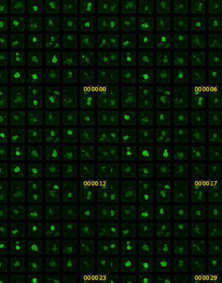
Image Credit: Yokogawa Life Science
- Nipkow spinning disk confocal technique enables high-speed but gentle 3D image acquisition
- Rich feature extraction for advanced cellular imaging analysis
- Wide field of view and tiling capabilities allow for simple imaging of large specimens
Enables analysis of time-lapse and live-cell
- The high precision stage incubator and low phototoxicity of the confocal allow for the examination of time-lapse and live-cell
- Max. 20fps option for fast time-lapse
High-quality image and similar operability to a traditional flow cytometer
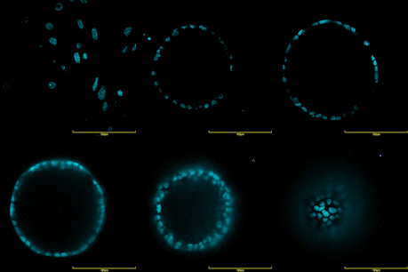
Image Credit: Yokogawa Life Science
- Unlike standard flow cytometry, removing cells from the culture dish is unnecessary
- Export data into FCS formats for easy downstream flow cytometry analyses
- Integration with the CellPathfiner software provides comprehensive analysis shown in real time with image acquisition
- Tracing back the data points is made simple with interactive graphs
Open platforms
- Connectable with external systems via handling robot
- Expandable to the integrated system as an image acquisition and quantification tool
- FCS/CSV/ICE data formats are readable by third-party data analysis tools
- A range of cell culture and sample dishes are applicable
Compact footprint, lightweight bench-top device; no need for a darkroom

*1 Option
*2 Contact to CQ1 partner for more information
Image Credit: Yokogawa Life Science
Multiple functions
Multiple functions fully integrated into a compact box
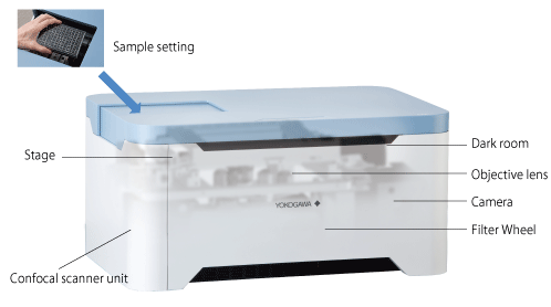
Image Credit: Yokogawa Life Science
The compact design has completely integrated functionality to provide an easy-to-use confocal imaging system without complex system integration. Users merely need to specify a sample and launch the software. A user-friendly interface and diverse functions help with measurement and analysis.
Principles of the microlens-enhanced Nipkow Disk Scanning Technology
A Nipkow spinning disk with about 20,000 pinholes and a subsidiary spinning disk with the same number of microlenses to concentrate excitation laser light into each matching pinhole are mechanically attached to a motor and rotate very quickly. This allows for a high-speed raster scan of the excitation lights on the material.
The patented design arranges the pinhole and microlenses on each disk to maximize the raster scan. Since multi-beam excitation only requires a small amount of laser power on the specimen to properly excite fluorescence, multi-beam scanning boosts scanning speed and considerably reduces photobleaching and phototoxicity.

Image Credit: Yokogawa Life Science
The CQ1 is the complete package for high-resolution, high content, high-quality analyses, with confocal quality images, reduced laser power usage and fast imaging speeds. With flexible image export options the CQ1 has the entire routine covered, from images to analysis.
Example of setup
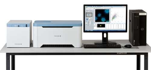
Image Credit: Yokogawa Life Science
Source: Yokogawa Life Science
| Item |
Specifications |
| Optics |
Microlens enhanced dual wide Nipkow disk confocal, Phase contrast (Optional add-on) |
| Laser/Filter |
Laser: Choose 2-4 lasers from 405/488/561/640 nm, 10-position Filter wheel (built-in) |
| Camera |
sCMOS 2560×2160 pixel, 16.6×14.0 mm |
| Objective lens |
Max.6 lenses (Dry: 2x, 4x, 10x, 20x, 40x Long working distance: 20x, 40x Phase contrast: 10x, 20x ) |
| Sample vessel |
Microplate (6, 24, 96, 384 well), Slide glass, Cover glass chamber*1, Dish (35, 60 mm*1) |
| XY stage |
High-precision XY stage, designated resolution 0.1 µm |
| Z focus |
Electric Z motor, designated resolution 0.1 µm |
| Autofocus |
Laser autofocus, Software autofocus |
| Feature data |
Number of cells/cellular granules, Intensity, Volume, Surface area, Area, Perimeter, Diameter, Sphericity, Circularity, etc |
| Data format |
Image: 16bit TIFF file (OME-TIFF), PNG
Numerical data: FCS, CSV, ICE |
| Workstation |
Measurement and analysis workstation |
| Size/weight |
Main unit: 600×400×298 mm 38 kg
Utility box: 275×432×298 mm 18 kg |
| Environment |
15 - 30 oC、20 - 70 %RH No condensation |
| Power consumption |
Main unit and Utility box: 100-240 VAC 800 VAmax, Workstation: 100-240 VAC 650 VAmax |
*1 Under development
*2 Display is not included with CQ1 system
Analysis software
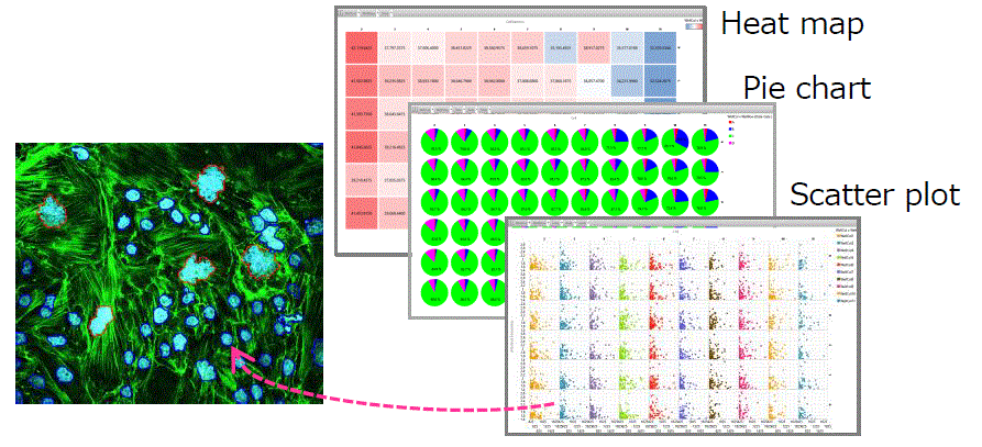
Image Credit: Yokogawa Life Science
CellPathfinder, high content analysis software (optional)
- Preset analysis menus with many application options
- Adaptable graph functions to show the findings of the analysis
- Direct link between chart and object image
Machine learning
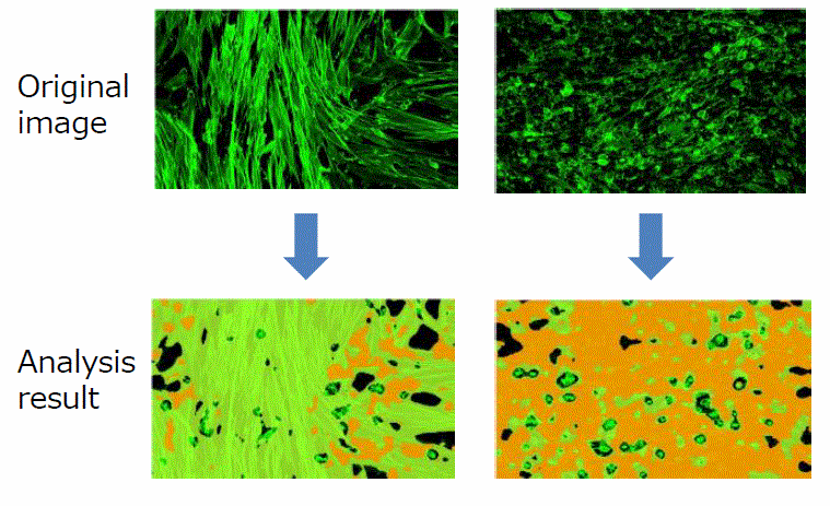 Image Credit: Yokogawa Life Science
The software learns the features of the sample objects that users have gathered.
3D analysis
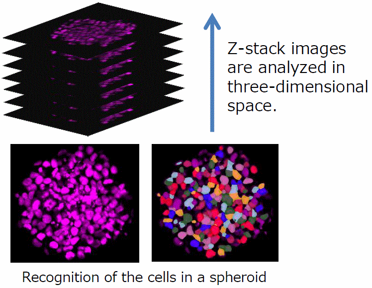
Image Credit: Yokogawa Life Science
Label-free analysis
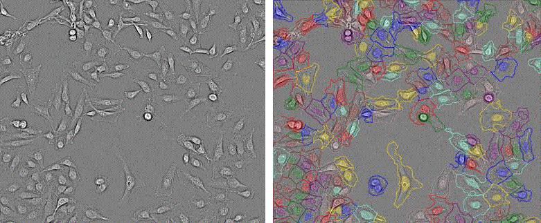
Image Credit: Yokogawa Life Science
The digital phase contrast function is an effective tool for unstained bright field sample analysis.
Compare CQ1, fluorescent imaging, and flow cytometry
Source: Yokogawa Life Science
| |
CQ1 |
General fluorescent imaging |
Flow cytometry |
| Cell removal/suspension treatment |
Not necessary |
Not necessary |
Necessary |
| Cell image confirmation |
Possible |
Possible |
Not possible |
| Display feature data and graphs in real-time with imaging |
Possible |
Depends on devices |
Possible |
| 3D data measurement |
Possible |
Not possible |
Not possible |
| Time-lapse |
Possible |
Not possible |
Not possible |
Software
CellActivision
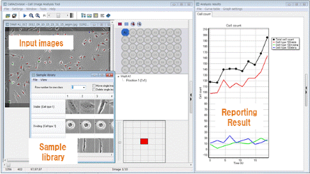
Image Credit: Yokogawa Life Science
CellActivision identifies cells or colonies directly from label-free images using Machine Learning Technology and a unique digital filter. CellActivision can also categorize and measure these cells using sample libraries created by the user.
CellPathfinder
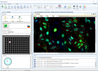
Image Credit: Yokogawa Life Science
Cell imaging software has a simplified, user-friendly interface, several graphing choices, and support for label-free samples.